Intelligent detection system for gynecology department
A detection system and gynecological technology, applied in the field of medical devices, can solve the problems of time-consuming and economical costs, patients losing treatment opportunities, etc., and achieve the effects of avoiding delays in disease, easy to carry, and convenient to use.
- Summary
- Abstract
- Description
- Claims
- Application Information
AI Technical Summary
Problems solved by technology
Method used
Image

Examples
Embodiment 1
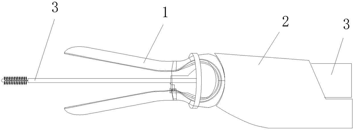
[0025] see Figure 1 ~ Figure 4 , the intelligent gynecological detection system of the present embodiment includes a vaginal speculum 1, an imaging mechanism 2, and a sampling brush 3. The vaginal speculum 1 is connected to the imaging mechanism 2, and the imaging mechanism 2 includes a main body 21, and the main body 21 is provided with a channel through which the sampling brush penetrates. Through the channel and vaginal dilator. In this embodiment, the sampling brush 3 is the detection mechanism.
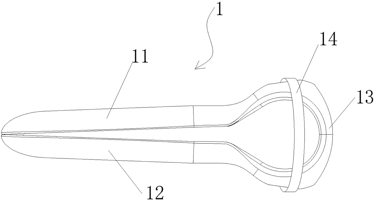
[0026] Such as figure 2 As shown, the vaginal dilator 1 includes an upper spreader 11 and a lower spreader 12, the upper spreader 11 and the lower spreader 12 are elastically connected by a bending part 13, and the bending part 13 is provided with a first An adjustment ring 14 for the opening of the opening angle between the expansion part and the second expansion part 12. The adjustment ring 14 moves back and forth on the outer ends of the first expansion part and the second...
Embodiment 2
[0035] see Figure 5 , the difference from Embodiment 1 is that in the intelligent gynecological detection system of this embodiment, the detection mechanism is the biopsy device 4, and the rest are the same as Embodiment 1, and will not be repeated here. The biopsy device 4 will be described in detail below.
[0036]The biopsy device 4 includes a sampling part, a connecting rod, and an operating handle. The sampling part comprises a protective cover 41 and a sampling tongs 42 arranged in the protective cover, the front end of the sampling part is provided with an opening (not shown) for the said sampling tongs to stretch out and retract, the connection between the sampling tongs 42 and the connecting rod 43 The front end is connected; there is a pressing device at the rear end of the operating handle 44, and the pressing device includes a clamping operation end (not shown) and a pressing head 45, one end of the pressing head 45 stretches out of the top of the operating handl...
Embodiment 3
[0039] see Figure 6 , The difference from Embodiment 1 is that the detection mechanism of the intelligent gynecology detection system in this embodiment is B-ultrasound mechanism 5, and the rest are the same as Embodiment 1, and will not be repeated here.
[0040] The B-ultrasound mechanism comprises a B-ultrasound probe 51, a connecting rod 52, a handle 53, and connecting wires (not shown in the figure). Probe connection; the handle is provided with a B-ultrasound data conversion module 54 for converting the signals obtained by the B-ultrasound probe, a battery 55 for power supply, and a PCB circuit board 56 for data processing and issuing control instructions. 56 is provided with a signal receiving end 57; the handle is provided with a data line 58 for connecting with an external display; the end of the handle is provided with a charging port 59 for connecting the battery to an external power source for charging.
[0041] In other embodiments, the data line 58 may not be p...
PUM
 Abstract
Description
Claims
Application Information
 - R&D
- Intellectual Property
- Life Sciences
- Materials
- Tech Scout
- Unparalleled Data Quality
- Higher Quality Content
- 60% Fewer Hallucinations
Browse by: Latest US Patents, China's latest patents, Technical Efficacy Thesaurus, Application Domain, Technology Topic, Popular Technical Reports.
© 2025 PatSnap. All rights reserved.Legal|Privacy policy|Modern Slavery Act Transparency Statement|Sitemap|About US| Contact US: help@patsnap.com