A cascaded fully convolutional neural network based ultrasound image segmentation method for thyroid nodules
A technology of convolutional neural network and thyroid nodules, which is applied in image semantic segmentation, based on the full convolutional neural network to automatically segment the nodule part of thyroid ultrasound images, which can solve low contrast, blurred borders of thyroid nodules, and calcification Point shadow accuracy and other issues
- Summary
- Abstract
- Description
- Claims
- Application Information
AI Technical Summary
Problems solved by technology
Method used
Image
Examples
Embodiment 1
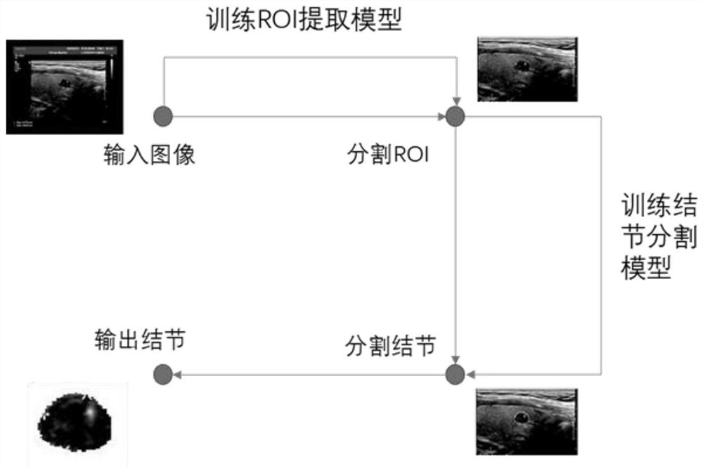
[0025] A cascaded fully convolutional neural network segmentation method for ultrasound images of thyroid nodules, see figure 1 , the method includes the following steps:
[0026] 101: Construct a simple fully convolutional neural network based on U-Net, segment the ultrasound image in the thyroid ultrasound data according to the simple fully convolutional neural network, and segment the region of interest from it;
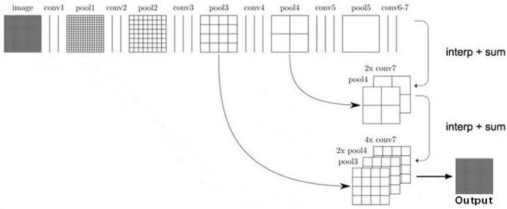
[0027] 102: Use the VGG19-FCN network as the downsampling layer to extract the deep features of the region of interest, so as to realize the automatic semantic segmentation of thyroid nodules;
[0028] Wherein, the simple and easy fully convolutional neural network in step 102 includes:
[0029] Five convolutional layers for downsampling, and five upsampling layers for upsampling;
[0030] Among them, the first five convolution conv are composed of two 3x3 convolution layers and one pooling layer, each convolution layer uses ReLU as the activation function, and ...
Embodiment 2
[0037] In order to achieve the above purpose, the technical idea of the embodiment of the present invention is: read the thyroid ultrasound data, remove the background area in the ultrasound image through a simple U-shaped fully convolutional neural network, construct training and test sample data sets, and construct a deep full volume The product neural network, namely FCN, is trained on the data set to obtain the semantic segmentation results suitable for ultrasound images. The specific steps include:
[0038] 201: Read the thyroid ultrasound data, and remove the background area of the ultrasound image through a simple full convolutional neural network based on U-Net:
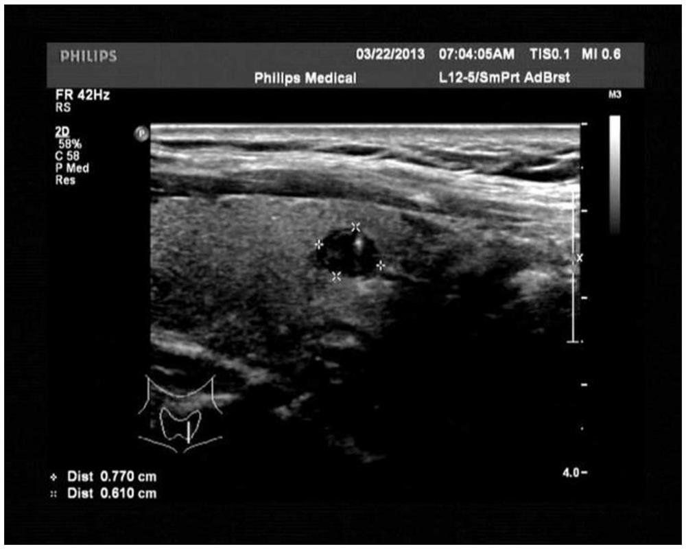
[0039] Among them, the ultrasound image is composed of a region of interest (ROI) and a background area. The ROI contains important diagnostic information, and the background area contains a large number of highlighted letters and symbols, which easily cause interference when the neural network extracts th...
Embodiment 3
[0067] Combine below Figure 1-Figure 3 The scheme in embodiment 1 and 2 is further introduced, see the following description for details:
[0068] 1) Read the ultrasound image data of thyroid nodules, which can be in various types of image formats:
[0069] 2) First read all the pictures in the training set, including ultrasound images and ROI-marked label data images, and train a UNET-based automatic ROI segmentation model (ie, the first part of the neural network);
[0070] 3) Using the segmented ROI and labeled data images of thyroid nodules under the guidance of experts as input, a model for automatic segmentation of thyroid nodules based on VGG19-FCN is trained, and the ROI segmentation model and nodule segmentation model are cascaded to form automatic segmentation The medical aid device for ultrasound images of thyroid nodules is then read into the test set data for testing. When using this method to automatically segment thyroid nodules, it is only necessary to read ...
PUM
 Abstract
Description
Claims
Application Information
 - R&D
- Intellectual Property
- Life Sciences
- Materials
- Tech Scout
- Unparalleled Data Quality
- Higher Quality Content
- 60% Fewer Hallucinations
Browse by: Latest US Patents, China's latest patents, Technical Efficacy Thesaurus, Application Domain, Technology Topic, Popular Technical Reports.
© 2025 PatSnap. All rights reserved.Legal|Privacy policy|Modern Slavery Act Transparency Statement|Sitemap|About US| Contact US: help@patsnap.com


