A method and apparatus for image analysis of aft-onset cataract

An image analysis and cataract technology, applied in the medical field, can solve the problems of time-consuming and energy-consuming, occupying the doctor's time and energy, unable to provide inspection results or analysis conclusions quickly and accurately, and achieve the effect of improving the acquisition efficiency.

Inactive Publication Date: 2018-12-28
ZHONGSHAN OPHTHALMIC CENT SUN YAT SEN UNIV
View PDF6 Cites 11 Cited by
  • Summary
  • Abstract
  • Description
  • Claims
  • Application Information

AI Technical Summary

Problems solved by technology

The method commonly adopted now is that people come to the hospital to see a doctor in person, and check in order according to the serial number, so that people spend a lot of time and energy on traffic and ranking; method, it will also take up a lot of time and energy of doctors
It can be seen that the current inspection method still cannot provide inspection results or analysis conclusions quickly and accurately.

Method used


Image

  • A method and apparatus for image analysis of aft-onset cataract
  • A method and apparatus for image analysis of aft-onset cataract
  • A method and apparatus for image analysis of aft-onset cataract

Examples


Embodiment 1

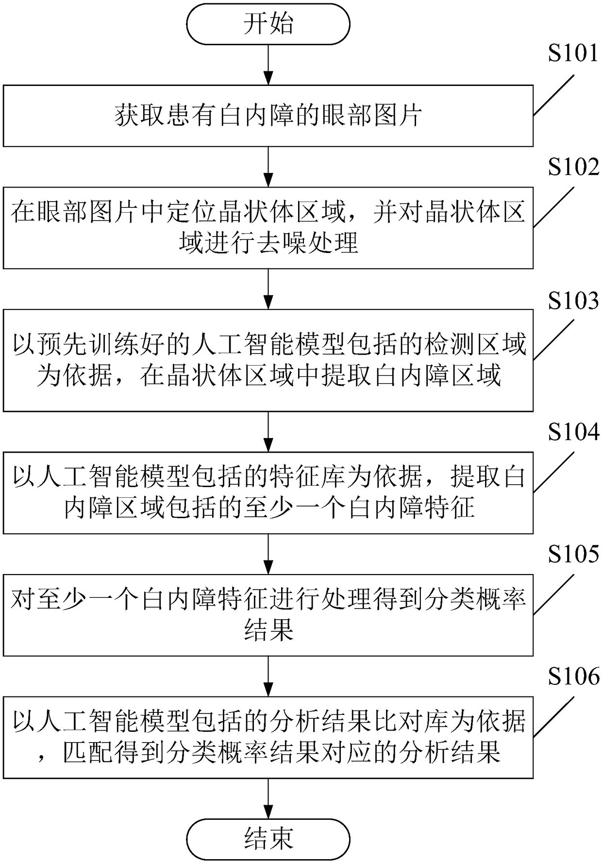
[0040] see figure 1 , figure 1 It is a schematic flowchart of an image analysis method for post-cataract provided in this embodiment.

[0041] Such as figure 1 As shown, this kind of after cataract image analysis method comprises the following steps:

[0042] S101. Obtain pictures of eyes with post-cataract.

[0043] As an optional implementation, obtaining pictures of eyes with post-cataract may include:

[0044] Receive a picture, determine whether the picture is an eye picture, if it is an eye picture, determine whether the eye picture is an eye picture with post-cataract, and if it is an eye picture with post-cataract, go to step S102.

[0045] Implementing this implementation method can accurately obtain eye pictures with post-cataract and avoid the mixing of eye pictures without post-cataract, thereby ensuring the matching degree between the pictures and the artificial intelligence model, thereby ensuring The stability and efficiency of the analytical method were co...

Embodiment 2

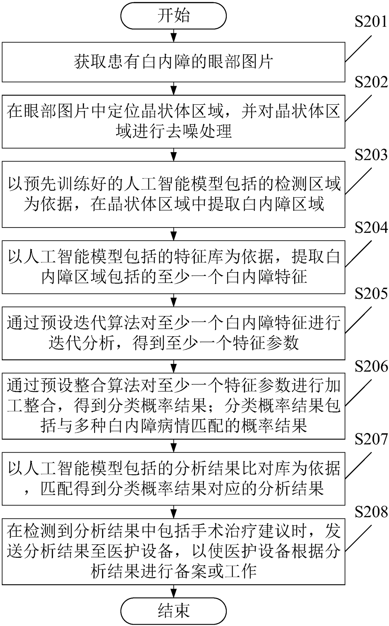
[0094] see figure 2 , figure 2 It is a schematic flowchart of an image analysis method for post-cataract provided in this embodiment. Such as figure 2 As shown, the post cataract image analysis method comprises the following steps:

[0095] S201. Obtain pictures of eyes with post-cataract.

[0096] Implementing this implementation manner can avoid receiving pictures of eyes with post-cataract that are not post-cataract information, and improve the processing efficiency of the artificial intelligence model.

[0097] S202. Locate the lens area in the eye image, and perform denoising processing on the lens area.

[0098] Implementing this embodiment, the pictures of eyes with post-cataract can be divided, so that the pictures of eyes with post-cataract can be divided into pictures of lens regions of post-cataract information. It can be seen that, by implementing this embodiment, the regional pictures including post-cataract information can be accurately obtained, and the ...

Embodiment 3

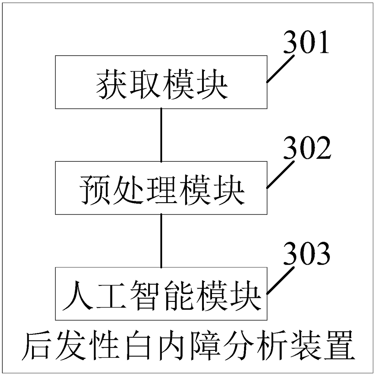
[0127] see image 3 , image 3 It is a schematic structural diagram of an after-cataract image analysis device provided by an embodiment of the present invention.

[0128] Such as image 3 As shown, the post-cataract image analysis device includes:

[0129] The acquiring module 301 is configured to acquire pictures of eyes with post-cataract.

[0130] The pre-processing module 302 is configured to locate the lens area in the eye picture and perform denoising processing on the lens area.

[0131] The artificial intelligence module 303 is configured to extract the post-cataract region in the lens region based on the pre-trained artificial intelligence model, and extract at least one post-cataract feature according to the post-cataract region.

[0132] The artificial intelligence module 303 is further configured to process at least one post-cataract feature based on the artificial intelligence model to obtain a classification probability result, so as to obtain an analysis re...


PUM


Abstract

The invention provides an image analysis method and a device for after-cataract, which comprises the following steps of: receiving an eye picture of after-cataract; a posterior capsule hyperplasia region of after-cataract is located and processed by slit-lamp posterior radiography; the area of posterior capsule proliferation is extracted from the area of posterior capsule proliferation according to the detection area included in the artificial intelligence model, wherein, the artificial intelligence model is pre-trained; at least one after-cataract feature included in the after-cataract regionis extracted based on the feature library included in the artificial intelligence model; at least one after cataract feature is processed to obtain a classification probability result; based on the analysis results comparison database included in the artificial intelligence model, matching results corresponding to the classification probability results are obtained. It can be seen that the methodmentioned above can simply, quickly and accurately determine the after-cataract of the patient, and give the corresponding analysis results to the user, so as to improve the efficiency of the user toseek medical treatment.

Description

technical field [0001] The invention relates to the medical field, in particular to an image analysis method and device for post-cataract. Background technique [0002] At present, people are paying more and more attention to medical care, and more and more people need to obtain their own health status to ensure the quality of work, study and life. However, in real life, it still takes a lot of time for people to obtain their health status, even when a large number of people are checking at the same time, the time people need to spend increases exponentially; on the other hand, when people do not need a comprehensive check Sometimes, the efficiency of the inspection will also be affected by matters such as registration and queuing, resulting in a waste of time and cost. It can be seen that how to provide people with fast and accurate inspection results or analysis conclusions has become a significant problem at present. [0003] Post-cataract, as a common complication, als...

Claims


Application Information

Patent Timeline
Patent Type & Authority Applications(China)
IPC IPC(8): G06T7/00G06K9/62H04L29/08
CPCH04L67/34G06T7/0012G06T2207/30041G06F18/2415
Inventor 林浩添龙尔平
Owner ZHONGSHAN OPHTHALMIC CENT SUN YAT SEN UNIV
Why Eureka
Try Eureka