
Nondestructive fluorescence detection spectrograph suitable for cell level

A fluorescence detection and cell-level technology, which is applied in the field of optical technology in the biological field, can solve the problems of small single cell size, inability to accurately excite cells, and accurate cell detection, so as to achieve great guiding significance and realize the effect of phosphorescence life test

Pending Publication Date: 2019-01-08
JIANGSU RAYME BIOTECH
View PDF0 Cites 1 Cited by
  • Summary
  • Abstract
  • Description
  • Claims
  • Application Information

AI Technical Summary

Problems solved by technology

However, the size of a single cell is very small, and the chemical components contained in it are also ultra-trace. However, the current analysis method Western blot needs to break millions of cells to achieve the detection of intracellular biochemical indicators. Therefore, traditional analysis Method is insufficient to detect differences at the level of individual cells
Not only that, the traditional methods are mostly fixed-point detection, which cannot realize real-time monitoring
[0003] The current fluorescence spectrometers are generally used for the detection of macroscopic samples, and there is no precise detection at the cell level. The main reason is that the diameter of the excitation light probe of the commonly used spectrometer is too thick, because the diameter of the general cell is 5-10 microns, and the diameter of the probe is usually It is difficult to accurately excite the cells from tens of millimeters to tens of centimeters. In addition, because the signal of the cells is relatively weak, it is difficult to control the laser power. If the power is too weak, the intracellular signals cannot be excited, and the power is too strong to cause damage to the cells.

Method used


Image

  • Nondestructive fluorescence detection spectrograph suitable for cell level
  • Nondestructive fluorescence detection spectrograph suitable for cell level

Examples


Embodiment Construction

[0011] The present invention will be further described below in conjunction with accompanying drawing.

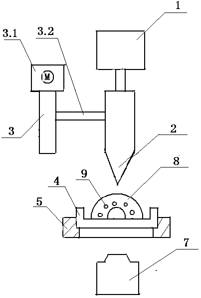
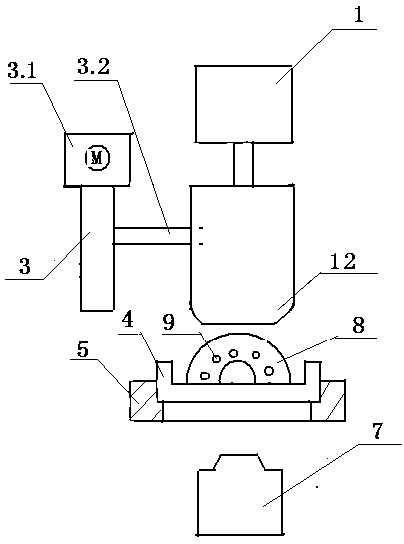
[0012] A spectrometer suitable for non-destructive fluorescence detection at the cell level, comprising an excitation light source 1, an excitation light probe 2 connected to the lower end of the excitation light source 1, the excitation light probe 2 is connected to the excitation light position control system 3 through the excitation light probe fixing device 3.2, and the excitation light The position control system 3 is equipped with a precision numerical control motor 3.1, and the excitation light probe 2 is provided with a sample pool 4 for placing cell samples. The cell sample 8 is provided with a fluorescent marker 9, and the fluorescence spectrum is collected and analyzed below the sample pool 4. System 7.

[0013] The excitation light probe fixing device 3.2 can drive the excitation light source 1 and the connected excitation light probe 2 to move up and down along...


PUM


Abstract

The invention discloses a cell level nondestructive fluorescence detection spectrograph. The cell level nondestructive fluorescence detection spectrograph comprises an exciting light source; an exciting light probe is connected to the lower end of the exciting light source; the exciting light probe is connected with an exciting light position control system through an exciting light probe fixing device; a precision NC motor is arranged on the exciting light position control system; a sample cell for placing cell samples is formed under the exciting light probe; and a fluorescence spectrum collecting and analyzing system is under the sample cell. The sample cell is formed a sample cell position control system which is hollowed-out in the middle and fixed on the surrounding. The cell level nondestructive fluorescence detection spectrograph disclosed by the invention can be used for single cell / sub-cell fluorescence analysis, can realize phosphorescence life testing and can detect the change of fluorescence intensity with time within a time scale range from milliseconds to hours; and the cell level nondestructive fluorescence detection spectrograph disclosed by the invention can be used for single cell / sub-cell fluorescence microscopy imaging, micro-area fluorescence spectrum analysis and integral steady-state fluorescence spectrum analysis in the X-Y direction and has greater guiding significance in early cancer detection and screening.

Description

technical field [0001] The invention relates to the technical field of application of optical technology in the biological field. Background technique [0002] In recent years, with the gradual deepening of research on cell physiology, scientists have discovered that even with the same genetic material and the same surrounding environment, these cells will still develop in different directions, and sometimes produce different functions. Individual cells appear to be the same, but their chemical composition and stress response are very different, and changes in individual cells are also related to major diseases such as cancer and neurological diseases. Single-celled biological processes in plants and animals are also far from fully understood. Single-cell analysis is therefore an important means of understanding and deciphering many critical life processes. However, the size of a single cell is very small, and the chemical components contained in it are also ultra-trace. H...

Claims


Application Information

Patent Timeline
IPC IPC(8): G01N21/64
CPCG01N21/64G01N21/6458G01N21/6486G01N2021/6417G01N2201/06113
Inventor 徐艇
Owner JIANGSU RAYME BIOTECH
Learn More