An automatic analysis and comparison method of fundus images and a storage device

A fundus image and automatic analysis technology, applied in the field of image processing, can solve the problems of heavy workload and low efficiency, and achieve the effect of improving efficiency, reducing workload and saving time

Active Publication Date: 2019-01-08
FUZHOU YIYING HEALTH TECH CO LTD
View PDF13 Cites 17 Cited by
  • Summary
  • Abstract
  • Description
  • Claims
  • Application Information

AI Technical Summary

Problems solved by technology

[0005] Therefore, it is necessary to provide an automatic fundus image analysis and comparison method to solve the problem that the prior art can only focus on the automatic analysis of a single image and the automatic detection of lesions, and the comparison of the fundus images is performed manually, which requires a large workload and high efficiency. low problem

Method used


Image

  • An automatic analysis and comparison method of fundus images and a storage device
  • An automatic analysis and comparison method of fundus images and a storage device
  • An automatic analysis and comparison method of fundus images and a storage device

Examples


Embodiment Construction

[0024] In order to explain in detail the technical content, structural features, achieved goals and effects of the technical solution, the following will be described in detail in conjunction with specific embodiments and accompanying drawings.

[0025] First, some nouns in this embodiment are explained as follows:

[0026] Optic disc: The full name is the optic disc, also called the optic nerve head. There is a light red disc-shaped structure with a diameter of about 1.5 mm and a clear boundary at about 3 mm from the macula to the nasal side of the retina. It is called the optic disc, or optic disc for short.

[0027] Macula: It is located 0.35cm below the temporal side of the optic disc in the fundus and slightly below it. It is in the optical center of the human eye and is the projection point of the visual axis.

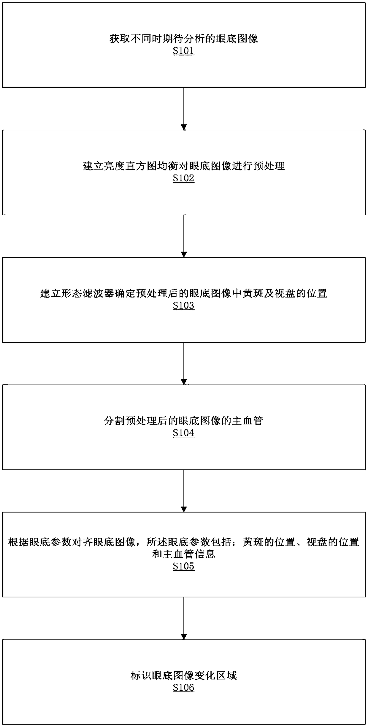
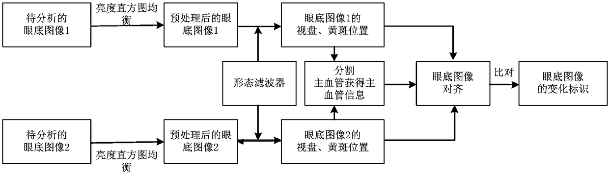
[0028] see Figure 1 to Figure 2 , in this embodiment, a method for automatic analysis and comparison of fundus images can be applied to a storage device, and t...


PUM


Abstract

The invention relates to the field of image processing, in particular to an automatic analysis and comparison method of fundus images. The automatic analysis and comparison method of fundus image comprises the following steps: fundus images expect to be analyzed at different times are obtained; the brightness histogram equalization is established to preprocess the fundus image; a morphological filter is established to determine the position of macula and optic disc in the preprocessed fundus image; segmentation of the main vessels of the preprocessed fundus image is carried out; the fundus image is aligned according to fundus parameters, and the fundus image change area is identified. By changing the area, a person can visualize the changes in the health of the eye fundus at different times, so as to quickly judge whether the eye fundus of an individual has health problems, obtain valuable information favorable for diagnosing diabetic retinopathy (DR), glaucoma and vascular changes, and assist in detecting and evaluating the diagnosis and treatment effect of the related chronic diseases, wherein the whole process does not require manual comparison and treatment., Thus, time is greatly saved, workload is reduced and efficiency is improved.

Description

technical field [0001] The invention relates to the field of image processing, in particular to an automatic fundus image analysis and comparison method and a storage device. Background technique [0002] The fundus is the only site in the human body where arteries, veins, and capillaries can be directly visualized non-invasively. By analyzing fundus images, valuable information can be obtained for diagnosing diabetic retinopathy (DR), screening for glaucoma, and vascular changes in hypertensive patients. A large number of studies and clinical practice have shown that regular fundus examination is the only effective way to detect lesions in time and obtain the best treatment opportunity, which has important guiding significance for the monitoring and prevention of various eye diseases and chronic diseases. [0003] The eyes are the only part of the body where blood vessels and nerves can be seen without surgery. Using a fundus camera to compare and observe the changes in t...

Claims


Application Information

Patent Timeline
Patent Type & Authority Applications(China)
IPC IPC(8): G06T7/00G16H50/20
CPCG06T7/0014G16H50/20G06T2207/30041G06T2207/30101G06T2207/20024
Inventor 余轮曹新容薛岚燕林嘉雯王丽纳
Owner FUZHOU YIYING HEALTH TECH CO LTD
Why Eureka
Try Eureka