A method and device for grading evaluation of brain glioma

A brain glioma, evaluation method technology, applied in the field of brain glioma classification evaluation method and device field, can solve the problems of inability to achieve classification evaluation, heavy workload, timely diagnosis of patients and hidden safety hazards of treatment, etc.

Active Publication Date: 2019-01-25
BEIJING PEREDOC TECH CO LTD
View PDF8 Cites 13 Cited by
  • Summary
  • Abstract
  • Description
  • Claims
  • Application Information

AI Technical Summary

Problems solved by technology

[0004] At present, for the grading evaluation of glioma, it is necessary to manually check and analyze the CT tomographic images one by one, especially the CT tomographic data of glioma is relatively large, and dozens or even hundreds of images can be obtained by sequential scanning. Therefore, for the grading and evaluation of glioma, the work

Method used


Image

  • A method and device for grading evaluation of brain glioma
  • A method and device for grading evaluation of brain glioma
  • A method and device for grading evaluation of brain glioma

Examples


Embodiment 1

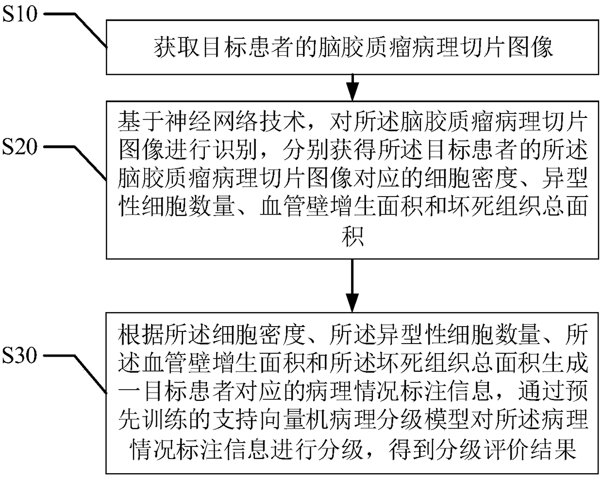
[0054] refer to figure 2 , the first embodiment of the present invention provides a method for grading evaluation of glioma, including:

[0055] Step S10, acquiring the pathological section image of the glioma of the target patient;

[0056] The above mentioned glioma pathological section image is a CT tomographic image of the patient's glioma.

[0057] Step S20, based on the neural network technology, identify the pathological slice image of the glioma, and respectively obtain the cell density, the number of atypia cells, and the hyperplasia of the vessel wall corresponding to the pathological slice image of the glioma of the target patient. area and total area of ​​necrotic tissue;

[0058] At present, manual grading and evaluation of glioma medical images is mainly judged from four aspects: cell density, cell atypia, vascular wall hyperplasia, and necrotic tissue.

[0059] Step S30, generate pathological condition labeling information corresponding to a target patient a...

no. 1 example

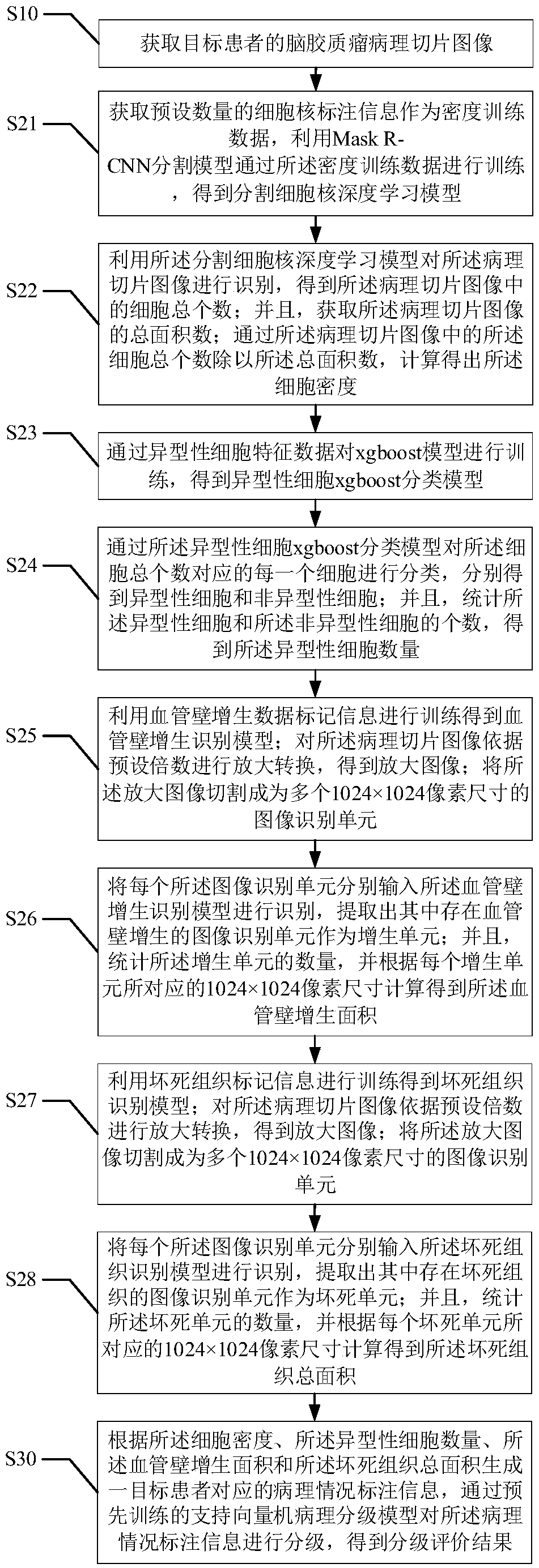
[0089] refer to image 3 , the second embodiment of the present invention provides a method for grading evaluation of glioma, based on the above figure 2 In the first embodiment shown, the obtaining of the cell density includes:

[0090] Step S21, obtaining a preset number of cell nucleus labeling information as density training data, using the Mask R-CNN segmentation model to train through the density training data, and obtaining a deep learning model for segmenting cell nuclei;

[0091] Step S22, using the segmented nucleus deep learning model to identify the pathological slice image to obtain the total number of cells in the pathological slice image; and obtain the total area of ​​the pathological slice image; The total number of cells in the slice image is divided by the total area to calculate the cell density.

[0092] As mentioned above, Mask R-CNN is a model that outputs high-quality instance segmentation masks while effectively detecting objects. It is an extensio...

Embodiment 3

[0123] refer to Figure 4 , the third embodiment of the present invention provides a method for grading evaluation of glioma, based on the above figure 2 In the first embodiment shown, after the "obtaining the graded evaluation result", it also includes:

[0124] Step S40, based on the image acquisition device, image acquisition is performed on the operating user who performs the grading evaluation, and an identity authentication image is obtained;

[0125] As mentioned above, the image acquisition device may be a camera, which is used for image acquisition and authentication of doctors.

[0126] Step S50, performing feature location, edge detection and threshold segmentation on the identity authentication image, and extracting facial features in the identity authentication image;

[0127] Step S60, using the pre-trained authentication user image recognition model to identify the facial features corresponding to the identity authentication image to determine whether the ope...


PUM


Abstract

The invention provides a method and device for grading evaluation of brain glioma, wherein the method comprises the following steps: obtaining a pathological slice image of the brain glioma of a target patient; Based on the neural network technology, the glioma pathological slice image is recognized, and the cell density, the number of atypical cells, the vascular wall hyperplasia area and the total necrotic tissue area corresponding to the glioma pathological slice image of the target patient are obtained respectively; and generating corresponding pathological condition labeling information of a target patient, and grading the pathological condition labeling information by a pre-trained support vector machine pathological grading model to obtain a grading evaluation result. The inventiongreatly improves the identification accuracy rate and the grading evaluation efficiency, reduces the workload of grading evaluation for the CT tomographic images of patients, and brings convenience tothe work of grading diagnosis.

Description

technical field [0001] The present invention relates to the technical field of tumor pathological grading, and more specifically, relates to a glioma grading evaluation method and a device thereof. Background technique [0002] Due to the influence of many factors, the global incidence of malignant tumors continues to rise. It is estimated that by 2020, the global incidence of malignant tumors will increase by 50%. Not only that, the number of deaths from malignant tumors is also rising rapidly around the world. In developing countries such as my country, this trend will be more obvious, and there will be a significant trend of younger people. Therefore, it is more urgent to strengthen the research on the prevention and treatment of malignant tumors, to evaluate the biological behavior and prognosis of tumors accurately and objectively, and to formulate treatment plans. [0003] Tumor type, grade and stage are currently the three most important indicators for evaluating tumo...

Claims


Application Information

Patent Timeline
IPC IPC(8): G06K9/00G06K9/62G06N3/08G06T7/00G06T7/11G06T7/13G06T7/136G16H30/20G16H40/67
CPCG06N3/08G06T7/0012G06T7/11G06T7/13G06T7/136G16H30/20G16H40/67G06T2207/30096G06V40/16G06F18/2411G06F18/214
Inventor 付钰王方
Owner BEIJING PEREDOC TECH CO LTD
Why Eureka
Try Eureka