
Coronary Artery Segmentation Method Fused with Dual Source CT Data

A technology of data and coronary artery, applied in the field of coronary artery segmentation fused with dual-source CT data, can solve the problem of data inconsistency

Active Publication Date: 2021-07-13
数坤(上海)医疗科技有限公司
View PDF4 Cites 0 Cited by
  • Summary
  • Abstract
  • Description
  • Claims
  • Application Information

AI Technical Summary

Problems solved by technology

[0005] However, there are challenges in its implementation: the data of the two phases are different, and the positions of the myocardium and blood vessels will change during the systolic and diastolic phases, and it is impossible to simply select high-quality images from one phase to replace another. Bad image in Phase 1

Method used


Image

  • Coronary Artery Segmentation Method Fused with Dual Source CT Data
  • Coronary Artery Segmentation Method Fused with Dual Source CT Data
  • Coronary Artery Segmentation Method Fused with Dual Source CT Data

Examples


Embodiment

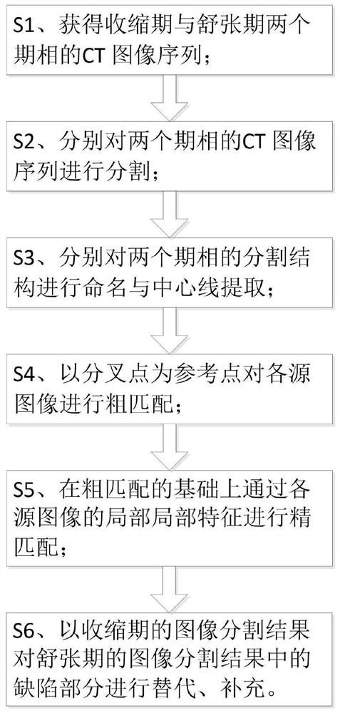
[0032] Please refer to figure 1 As shown, the present invention discloses a coronary artery segmentation method that fuses dual-source CT data, including:

[0033] S1. Perform a dual-source CT scan to obtain CT image sequences in the systolic and diastolic phases;

[0034] S2. Segmenting the CT image sequences of systole and diastole respectively;

[0035] S3. Name each blood vessel in the segmentation result of the systolic and diastolic CT image sequences and extract the centerline.

[0036] Wherein, the naming of each blood vessel is to facilitate subsequent establishment of a mapping relationship, and the center line is used as a basis for establishing the mapping relationship.

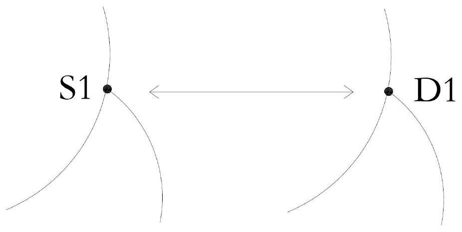
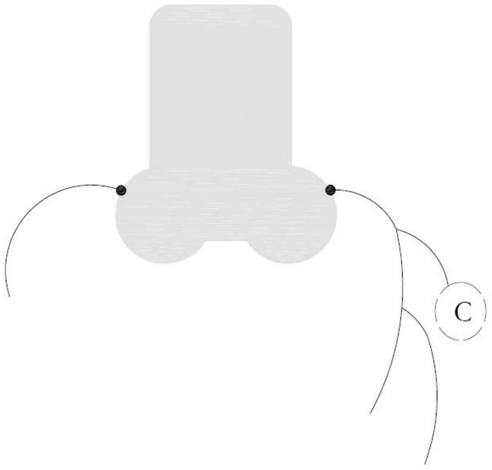
[0037] S4. Search for bifurcation points in each centerline, and perform rough matching on each source image with the bifurcation point as a reference point.

[0038] For 3D data matching, theoretically, the more control points (reference points) are provided, the more accurate the matching is,...


PUM


Abstract

The invention discloses a method for coronary artery segmentation fused with dual-source CT data, comprising: S1, performing dual-source CT scans, respectively obtaining CT image sequences of the systolic phase and the diastolic phase; S2, respectively analyzing the CT images of the two phases sequence; S3, name each blood vessel in the segmentation results of the two phase CT image sequences and extract the centerline; S4, find the bifurcation point in each centerline, and use the bifurcation point as a reference point to Perform rough matching on the CT images of the two phases; S5, extract local matching features in the CT image sequences of the two phases through the neural network, perform fine matching on the basis of rough matching through the local matching features, and obtain two The mapping relationship of the CT image sequence of phase; S6, based on the CT image sequence and segmentation result of the diastolic period, select the corresponding part from the CT image of the systolic period and the segmentation result to splice or replace the defective part of the image, and output Fused CT image sequence and segmentation results.

Description

technical field [0001] The invention relates to the technical field of CT image post-processing, in particular to a coronary artery segmentation method for fusing dual-source CT data. Background technique [0002] CT has been used in clinical examination soon after its birth, especially after the emergence of spiral CT, it has been widely used in the examination and diagnosis of various parts of the human body. Since the traditional spiral CT has only one set of X-ray generating device and one set of detector system, when scanning high-speed moving objects (such as coronary arteries, one inspection must be completed within a limited time, and it is necessary to ensure that the patient is as safe as possible during the scan. No breathing movement, otherwise, blurred images and jagged artifacts will appear in mild cases, and no diagnostic images can be obtained at all in severe cases, and the examination cannot be completed) will appear powerless. [0003] In this regard, Sie...

Claims


Application Information

Patent Timeline
Patent Type & Authority Patents(China)
IPC IPC(8): G06T5/50G06K9/34G06K9/46
CPCG06T5/50G06T2207/30168G06V10/267G06V10/44
Inventor 肖月庭阳光郑超
Owner 数坤(上海)医疗科技有限公司
Learn More