A lung nodule medical image registration method and a device thereof,

A technology for medical imaging and pulmonary nodules, applied in the field of image recognition, can solve the problems of inaccurate classification, reduced overall registration efficiency, and long registration time.

Active Publication Date: 2019-02-22
BEIJING PEREDOC TECH CO LTD
View PDF7 Cites 12 Cited by
  • Summary
  • Abstract
  • Description
  • Claims
  • Application Information

AI Technical Summary

Problems solved by technology

[0003] Existing medical image registration methods all directly recognize the obtained medical images of patients. There is a lot of noise in the recognition target image, which leads to a long registration time and a large amount of data calculation in the registration image. , greatly reducing the overall registration efficiency
In addition, because pulmonary nodules are in medical imaging, in the cross-sectional tomograph

Method used


Image

  • A lung nodule medical image registration method and a device thereof,
  • A lung nodule medical image registration method and a device thereof,
  • A lung nodule medical image registration method and a device thereof,

Examples


Embodiment 1

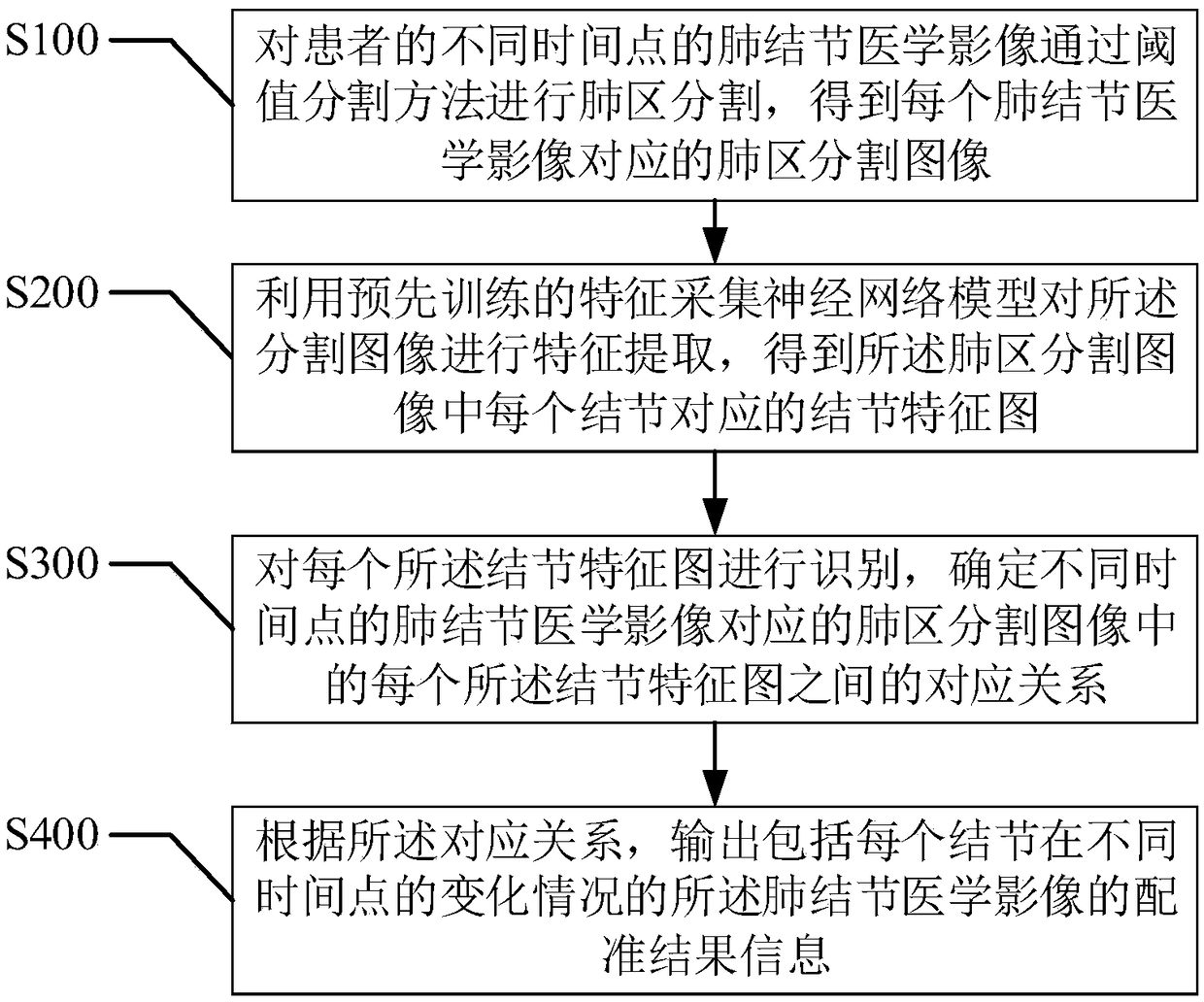
[0055] refer to figure 2 , the first embodiment of the present invention provides a medical image registration method for pulmonary nodules, including:

[0056] Step S100 , performing lung region segmentation on the medical images of pulmonary nodules at different time points of the patient through a threshold segmentation method to obtain a lung region segmentation image corresponding to each pulmonary nodule medical image;

[0057] As mentioned above, the medical image of pulmonary nodules refers to the medical image obtained from the CT scan of the lungs of the patient, which may contain pulmonary nodules or other tissues. In this embodiment, the target is a tomographic image of the lungs.

[0058] It should be noted that CT (Computed Tomography), that is, electronic computerized tomography, uses precisely collimated X-ray beams, γ-rays, ultrasonic waves, etc., together with highly sensitive detectors to make a joint around a certain part of the human body. A cross-secti...

Embodiment 2

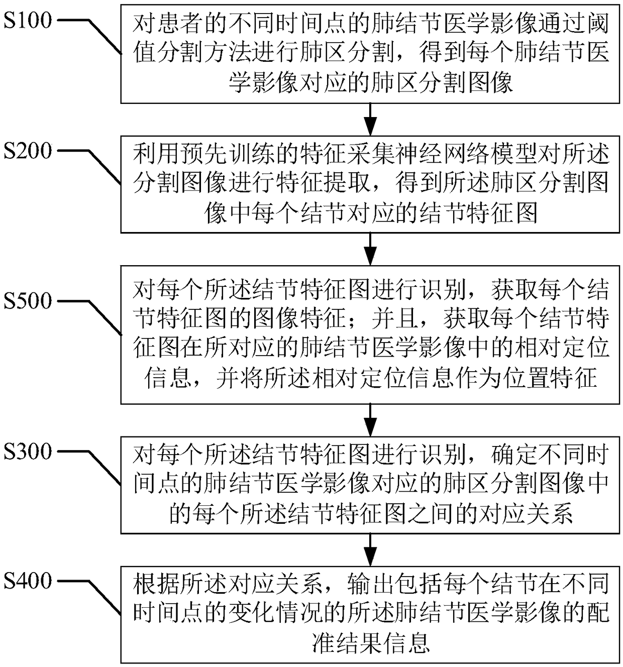
[0076] refer to image 3 , the second embodiment of the present invention provides a medical image registration method for pulmonary nodules, based on the above figure 2 In the first embodiment shown, the step S300, "recognize each nodule feature map, and determine each nodule in the lung region segmentation image corresponding to the pulmonary nodule medical image at different time points Before the correspondence between feature maps", it also includes:

[0077] Step S500, identifying each of the nodule feature maps, obtaining the image features of each nodule feature map; and obtaining the relative positioning information of each nodule feature map in the corresponding pulmonary nodule medical image, And use the relative positioning information as a position feature.

[0078] As mentioned above, each nodule feature map corresponds to an image feature obtained through recognition, and each nodule feature map has a corresponding location information in the original pulmona...

Embodiment 3

[0081] refer to Figure 4-5 , the third embodiment of the present invention provides a medical image registration method for pulmonary nodules, based on the above image 3 In the second embodiment shown, in the step S500, "recognize each of the nodule feature maps, and determine the corresponding lung nodule medical images at different time points according to the feature information of each of the nodule feature maps. The corresponding relationship between each of the nodule feature maps in the lung region segmentation image" includes:

[0082] Step S510, taking every two nodule feature maps as a recognition group, and extracting two nodule feature maps in all the recognition groups to match each other according to the image features corresponding to each nodule feature map The recognition group of is used as an image recognition association group;

[0083] As mentioned above, the medical images of pulmonary nodules at different time points can contain a certain number of n...


PUM


Abstract

The invention provides a lung nodule medical image registration method and a device thereof, wherein the method comprises the following steps of segmenting the lung regions of the lung nodule medicalimages of patients at different time points through a threshold segmentation method to obtain segmented images of the lung regions; using a feature collection neural network model for feature extraction of the segmented image to obtain a nodule feature map corresponding to each nodule; identifying each nodule feature map and determining the corresponding relationship between each nodule feature map at different time points; according to the correspondence, outputting the registration result information. The method provided by the invention is based on neural network technology, has simple algorithm and high identification efficiency, greatly reduces the registration time, and can realize the accurate registration of nodules in lung images, and improves accuracy rate.

Description

technical field [0001] The present invention relates to the technical field of image recognition, and more specifically, to a medical image registration method and device for pulmonary nodules. Background technique [0002] When doing medical image analysis, it is often necessary to analyze several images of the same patient together, so as to obtain comprehensive information of the patient and improve the level of medical diagnosis and treatment. For quantitative analysis of several different images, the strict alignment of these images must first be solved, which is the so-called image registration. Medical image registration refers to seeking a kind of (or a series of) spatial transformation for a medical image to make it consistent with the corresponding points on another medical image. This consistency means that the same anatomical point on the human body has the same spatial position in the two matching images. The result of registration should match all anatomical ...

Claims


Application Information

Patent Timeline
IPC IPC(8): G06T7/33G06T7/136
Inventor 付钰胡飞
Owner BEIJING PEREDOC TECH CO LTD
Why Eureka
Try Eureka