Image report based CT image emphysema automatic labeling method

A CT image, automatic labeling technology, applied in medical images, instruments, characters and pattern recognition, etc., can solve the problem of unmarked emphysema location and area size, no emphysema quantitative analysis results, no lesion location information, etc. , to reduce the cost of seeing a doctor, save the time of diagnosis, and improve the efficiency of diagnosis

Inactive Publication Date: 2019-03-08
沈阳医学院附属中心医院
View PDF8 Cites 12 Cited by
  • Summary
  • Abstract
  • Description
  • Claims
  • Application Information

AI Technical Summary

Problems solved by technology

[0004] At present, there are a large number of image reports and CT image data for hospitals, but information such as the location and size of the emphysema is not marked on the CT images
[0005] A large amount of medical data: At present, there are CT images and corresponding imaging diagnosis reports of a certain scale of COPD patients in the hospital, but doctors have not marked the location of emphysema on the CT images, and there is no quantitative analysis result of emphysema. Only qualitative text description of emphysema, no specific location information of lesion

Method used


Image

  • Image report based CT image emphysema automatic labeling method
  • Image report based CT image emphysema automatic labeling method
  • Image report based CT image emphysema automatic labeling method

Examples


Embodiment Construction

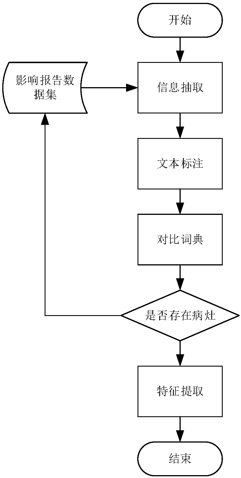
[0038] The present invention will be further described below in conjunction with accompanying drawing. It should be understood that the preferred embodiments described here are only used to illustrate and explain the present invention, not to limit the present invention.

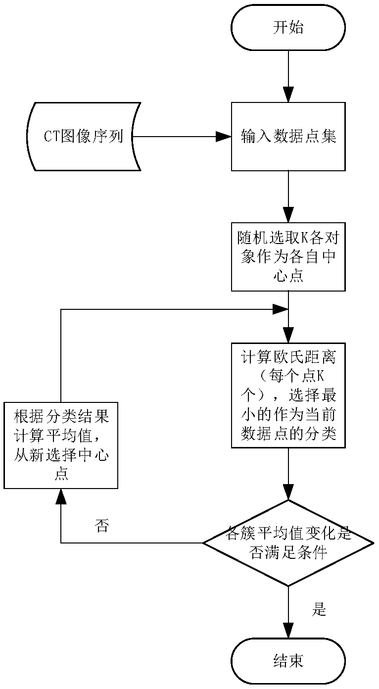
[0039] As shown in the figure, the method for automatically labeling emphysema in CT images based on image reports, the steps are as follows: (1), complete the image report report, CT image sequence input and image standardization preprocessing in the input module; (2), in the input module In the speech semantic extraction module, the feature information in the image report is extracted according to the dictionary technology and the rule pattern matching technology, that is, the information about emphysema description; (3), in the emphysema lesion extraction module, the lungs are first divided into regions , the width of each area of ​​the right lung from top to bottom is the same, and the left lung will be ...


PUM


Abstract

The invention relates to an image report based CT image emphysema automatic labeling method. The method includes the following steps: (1) accomplishing an image report, CT image sequence inputting andimage standardization preprocessing in an input module; (2) extracting the characteristic information, namely the information about emphysema description, in the image report in a phonetic and semantic extraction module according to the technical and rule pattern matching technology of a dictionary; (3) performing regional division on a lung in an emphysema focus extraction module, and performingcluster analysis in a lung area to extract emphysema focus; and (4) obtaining the CT threshold value of each area through calculation according to calculating results of the step 3, labelling the emphysema area of each area in an output and display module, displaying the emphysema areas in a CT image, and giving a quantitative analysis report of lung function. The advantages of the method are that processes during emphysema diagnosis and treatment can be simplified, and medical expenses of patients can be reduced.

Description

technical field [0001] The invention relates to an automatic labeling method for CT image emphysema, in particular to an automatic labeling method for CT image emphysema based on an image report. Background technique [0002] Chronic obstructive pulmonary disease (COPD), referred to as COPD, is a disease that seriously threatens human health. At present, the main means for doctors to diagnose COPD rely on pulmonary function tests and CT imaging scans, but the imaging reports do not indicate The location information of emphysema on CT images, and pulmonary function tests cannot provide information on the location of emphysema lesions, which leads to the quantitative evaluation and severity grading of emphysema relying on subjective experience, which affects the early diagnosis of patients. Prone to missed diagnosis and misdiagnosis, reducing the effectiveness of diagnosis. [0003] At present, the post-processing workstations equipped by some CT manufacturers provide manual ...

Claims


Application Information

Patent Timeline
Patent Type & Authority Applications(China)
IPC IPC(8): G16H30/20G16H30/40G06K9/62
CPCG16H30/20G16H30/40G06F18/23213
Inventor 夏书月康雁何巍孙龙昊李强陈磊
Owner 沈阳医学院附属中心医院
Why Eureka
Try Eureka