A biosensor for detecting uracil glycosidase (UDG) activity and its preparation method

A technology of uracil glycosidase and detection method, which is applied in the direction of instruments, measuring devices, scientific instruments, etc., and can solve the problems of easy bleaching of fluorescence detection and low accuracy of colorimetric detection

Active Publication Date: 2021-06-15
UNIV OF JINAN
View PDF5 Cites 0 Cited by
  • Summary
  • Abstract
  • Description
  • Claims
  • Application Information

AI Technical Summary

Problems solved by technology

[0006] Aiming at the problems of low precision of colorimetric detection and easy bleaching of fluorescence detection, the present invention provides a biosensor for detecting UDG activity, which adopts surface-enhanced Raman scattering (SERS) technology to achieve ultra-sensitive and precise detection. At the same time, the present invention also provides the biosensor Preparation method of sensor

Method used


Image

  • A biosensor for detecting uracil glycosidase (UDG) activity and its preparation method
  • A biosensor for detecting uracil glycosidase (UDG) activity and its preparation method
  • A biosensor for detecting uracil glycosidase (UDG) activity and its preparation method

Examples


Embodiment 1

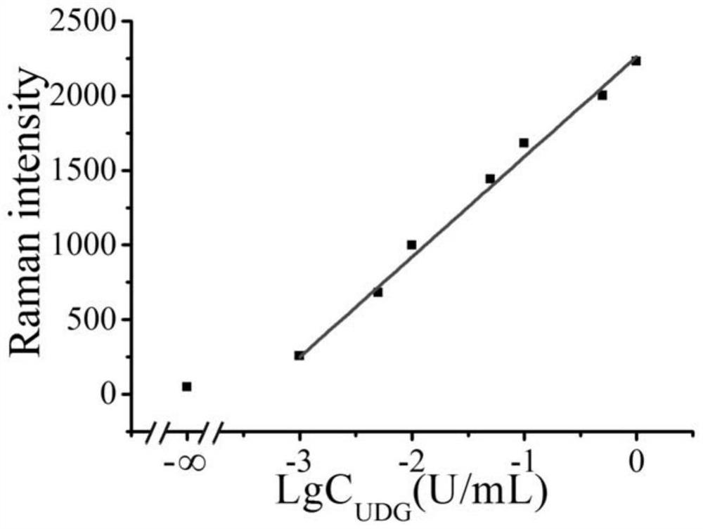
[0043] Example 1 Pure UDG concentration detection

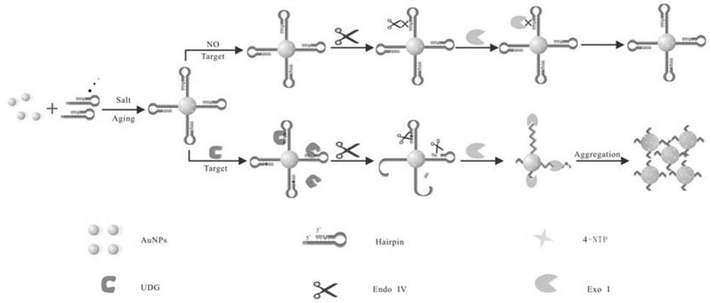
[0044] 1. Modification of Raman dye and hairpin probe DNA onto the surface of gold nanoparticles:

[0045] a. Take 1 mL nano-gold solution in a centrifuge tube, centrifuge for 10 min, and centrifuge the two tubes at the same time for later use. Centrifuge until the supernatant is colorless and transparent, remove the supernatant, and add 300 μL of sterile water to concentrate the nano-gold solution to 3 nM. Transfer to a 1 mL glass vial, seal it with tinfoil, and add 12 μL of Raman dye (4-NTP) with a concentration of 0.25 nM.

[0046] b. After standing at room temperature for 30 min, add 150 μL of -SH-modified substrate probe (hairpin DNA) with a concentration of 30 μM, mix well, and place at 4 °C for 24 h.

[0047] c. Slowly add 50 μL of PB buffer solution (100 mM conventional phosphate buffer solution, pH=7.4) several times, add magnets (soaked in aqua regia the day before) and stir for 10 min, then continue to add 27 μL ...

Embodiment 2

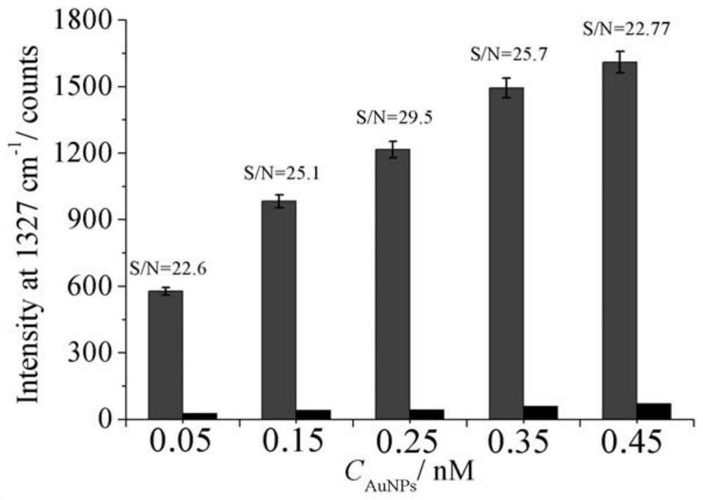
[0052] Embodiment 2 optimizes Raman dye concentration to improve signal-to-background ratio

[0053] 1. The steps for modifying Raman dyes and hairpin probes (-SH) to the surface of gold nanoparticles are as follows:

[0054] a. Take 1 mL nano-gold solution in a centrifuge tube, centrifuge for 10 min, and centrifuge the two tubes at the same time for later use. Centrifuge until the supernatant is colorless and transparent, remove the supernatant, and add 300 μL of sterile water to concentrate the nano-gold solution to 3 nM. Transfer to a 1 mL glass bottle, seal it with tin foil, and add 12 μL of different concentrations of Raman dyes (4-NTP).

[0055] b. After standing at room temperature for 30 min, add 150 μL of hairpin probe modified with -SH with a concentration of 30 μM, mix well, and place at 4 °C for 24 h.

[0056] c. Slowly add 50 μL of PB buffer several times, add magnets (soaked in aqua regia the day before) and stir for 10 min, then continue to add 27 μL of PBS bu...

Embodiment 3

[0062] The UDG concentration in the mixed solution of embodiment 3 actual measurement

[0063] 1. Steps for modifying Raman dyes and hairpin probes (-SH) onto the surface of gold nanoparticles:

[0064] Same as in Example 1, the hairpin probe and Raman dye were modified onto the surface of gold nanoparticles.

[0065] 2. Measure the concentration of UDG enzyme in the mixture

[0066] a. Mix the labeled gold nanoparticles solution (12 μL), endonuclease IV (2 U / mL), 10×NEBuffer (10 mMTris-HCl, 50 μM NaCl, 10 mM MgCl 2 , 1 mM DTT, pH 7.9) 2 uL, water, and crude cell extract were added to centrifuge tubes, shaken for 30 s, and placed in a water bath at 37 °C for 60 min.

[0067] b. After 60 minutes, take out the mixed solution from the water bath, observe the color change, and detect the peak intensity with a Raman spectrometer, and detect the target object accordingly.

[0068] The test results are shown in the table below. It can be seen that when the concentration of UDG is ...


PUM

PropertyMeasurementUnit
molecular weightaaaaaaaaaa

Abstract

The invention relates to the technical field of biosensors, in particular to a biosensor for detecting uracil glycosidase activity based on the surface-enhanced Raman scattering effect generated by the agglomeration of gold nanoparticles, and a preparation method thereof. Aiming at the problems of low precision of colorimetric detection and easy bleaching of fluorescence detection, the present invention realizes fast, sensitive and safe detection of UDG enzyme activity by using hairpin nucleic acid probes modified on the surface of gold nanoparticles as substrates, and using surface-enhanced Raman Scattering (SERS) technology realizes ultra-sensitive and precise detection, and the invention also provides a preparation method of the biosensor, which is carried out in a homogeneous solution with mild conditions and easy operation.

Description

technical field [0001] The invention relates to the technical field of biosensors, in particular to a biosensor for detecting uracil glycosidase activity based on the surface-enhanced Raman scattering effect generated by the agglomeration of gold nanoparticles, and a preparation method thereof. Background technique [0002] Since Lindahl et al first reported the activity of UDG (uracil glycosidase) in Escherichia coli extracts in 1974, [0003] UDG research is getting more and more attention. UDG is a monomeric protein with a molecular weight of about 25Kda and widely exists in various prokaryotes and eukaryotes. As the main member of base damage repair protein, UDG can specifically recognize DNA [0004] Uracil residues in single or double strands can hydrolyze the N-glycosidic bond between it and deoxyribose, thereby removing uracil and generating an abasic site (AP site). After endonuclease IV and exonuclease III recognize the AP site, they cut off the phosphodiester b...

Claims


Application Information

Patent Timeline
Patent Type & Authority Patents(China)
IPC IPC(8): G01N21/65
CPCG01N21/658
Inventor 王玉王海旺黄加栋刘素王敬锋张雪宋晓蕾
Owner UNIV OF JINAN
Why Eureka
Try Eureka