3D printed occlusal splint for the treatment of temporomandibular joint disorders and its preparation method

A temporomandibular joint and 3D printing technology, which is applied in the field of oral cavity and medical equipment, can solve the problems of single design of treatment equipment, complex treatment procedures, and poor accuracy of equipment, and achieve a highly targeted, high-quality, simple and convenient use Effect

Active Publication Date: 2020-09-22
GENERAL HOSPITAL OF PLA
View PDF7 Cites 0 Cited by
  • Summary
  • Abstract
  • Description
  • Claims
  • Application Information

AI Technical Summary

Problems solved by technology

[0006] In order to overcome the single design of treatment equipment for temporomandibular joint disorders in the prior art, resulting in complicated treatment procedures, poor prognosis due to poor precision of equipment, etc., the present invention provides a 3D digital technology combined with 3D printing technology. Printed occlusal splint for treating temporomandibular joint disorder and preparation method thereof

Method used


Image

  • 3D printed occlusal splint for the treatment of temporomandibular joint disorders and its preparation method
  • 3D printed occlusal splint for the treatment of temporomandibular joint disorders and its preparation method
  • 3D printed occlusal splint for the treatment of temporomandibular joint disorders and its preparation method

Examples


Embodiment 1

[0066] Example 1 A preparation method of a 3D printed occlusal splint for the treatment of temporomandibular joint disorders

[0067] A method for preparing a 3D printed occlusal splint for treating temporomandibular joint disorders, specifically:

[0068] (1) Data Acquisition and Processing

[0069] 1. Cone beam CT was used to shoot the patient's craniomaxillofacial; cone beam CT applied DICOM3.0 format, and the slice thickness was set to 1.25mm; the shooting range was from the supraorbital rim to the mandibular rim.

[0070] 2. Use a 3D scanner to scan the data of the maxillary dentition, mandibular dentition, and occlusion of the upper and lower dentition; use a 3D scanner to scan the data of the upper dentition, mandibular dentition, and occlusion of the upper and lower dentition. The range is: the scanning range of the maxillary dentition is the upper dentition including at least the entire tooth surface of two pairs of symmetrical molars and the upper palate; the scanni...

Embodiment 2

[0080] Example 2 A preparation method of a 3D printed occlusal splint for the treatment of temporomandibular joint disorders

[0081] A method for preparing a 3D printed occlusal splint for treating temporomandibular joint disorders, specifically:

[0082] (1) Data Acquisition and Processing

[0083] 3. Use cone beam CT to shoot the patient's craniomaxillofacial; cone beam CT uses DICOM3.0 format, and the slice thickness is set to 1.25mm; the shooting range is from the upper orbital edge to the lower edge of the mandible

[0084] 4. Use a 3D scanner to scan the data of the maxillary dentition, mandibular dentition, and occlusal dentition; use a 3D scanner to scan the data of the upper dentition, mandibular dentition, and occlusion of the upper and lower dentition. The range is: the scanning range of the mandibular dentition is the entire tooth surface of the teeth corresponding to the upper dentition; the scanning range of the upper and lower dentition is the outer surface of...

Embodiment 3

[0096] Example 3 A preparation method of a 3D printed occlusal splint for the treatment of temporomandibular joint disorders

[0097] A method for preparing a 3D printed occlusal splint for treating temporomandibular joint disorders, specifically:

[0098] (1) Data Acquisition and Processing

[0099]5. Use cone-beam CT to shoot the patient's craniomaxillofacial; cone-beam CT uses DICOM3.0 format, and the slice thickness is set to 1.25mm; the shooting range is from the upper orbital edge to the lower edge of the mandible

[0100] 6. Use a three-dimensional scanner to scan the data of the maxillary dentition, mandibular dentition, and occlusal dentition respectively; use a 3D scanner to scan the data of the upper dentition, mandibular dentition, and occlusion of the upper and lower dentition respectively The scope is: when the upper and lower dentition are occlusal, the scanning range is the outer surface of the upper and lower dentition; the 3D scanner is Smartoptics, 3shape o...


PUM

PropertyMeasurementUnit
thicknessaaaaaaaaaa
thicknessaaaaaaaaaa
thicknessaaaaaaaaaa

Abstract

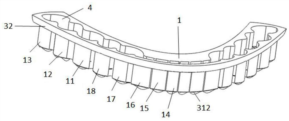
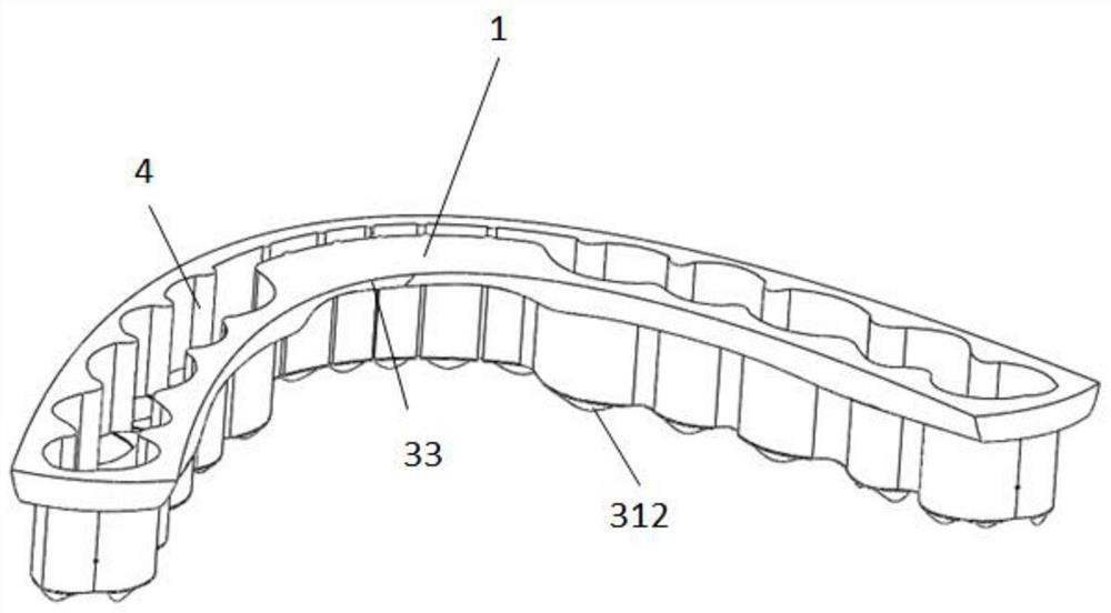
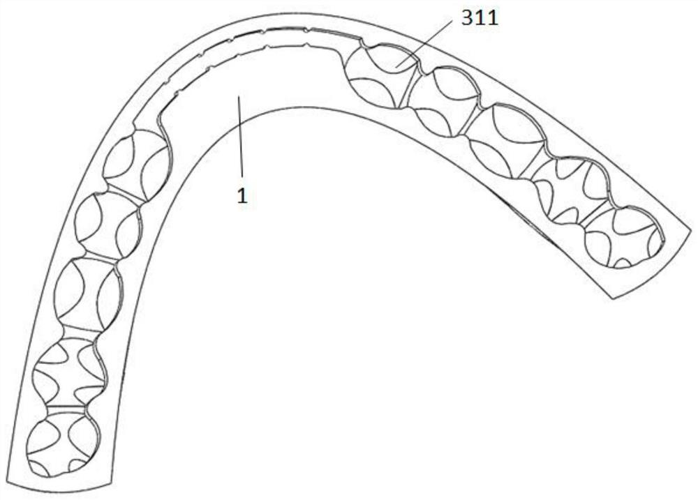
The invention discloses a biteplate for treating temporomandibular disorders by 3D printing and a preparation method thereof to solve the problem that the biteplate in treating temporomandibular disorders is simplex in design, complex in flow of the treatment process and imprecise to manufacture. The preparation method specifically comprises the following steps: scanning craniomaxilloface by spinal canal bundle CT; acquiring dentition data of upper jaw dentition, low jaw dentition and occlusion of upper and lower jaw dentitions; then importing the data into Super Virtual software to reconstruct upper and low jaws and dentitions; upshifting the upper jaw dentition data; and importing the upshifted and original upper jaw dentition data into CAD for designing the biteplate and printing the same. The biteplate comprises tooth occlusion sleeves of at least two pairs of symmetrically motar teeth, the tooth occlusion sleeves are connected through connecting plates, and teeth placing holes arecorrespondingly formed in the tooth occlusion sleeves; the connecting plates are arranged on the inner and outer sides, the shape of the inner bottom surface of each tooth occlusion sleeve is consistent to that of the bottom surface of the teeth of the upper jaw dentition, the bottom surface of the outer side of each tooth occlusion sleeve is engaged to the lower jaw dentition effectively, and the tooth occlusion sleeves are connected in a penetrating manner.

Description

technical field [0001] The invention belongs to the technical field of medical devices used in the field of oral cavity, in particular to a treatment device for temporomandibular joint disorders, in particular to a 3D printed occlusal splint for treating temporomandibular joint disorders and a preparation method thereof. Background technique [0002] Temporomandibular disorders (TMD) are common clinical diseases in stomatology, and the main clinical manifestations are local joint soreness or pain, joint snapping and mandibular movement disorder. Pain can be in the joint area or around the joint; it can be accompanied by tenderness of varying severity; joint soreness or pain is especially obvious when chewing and opening the mouth, and the snapping sound occurs when the mouth is opened, and the sound can occur at different stages of jaw movement , which can be a crisp single sound or a broken continuous sound. Common movement obstacles are limited mouth opening, deviation of ...

Claims


Application Information

Patent Timeline
Patent Type & Authority Patents(China)
IPC IPC(8): A61C19/00A61F5/00B29C64/386B29C64/10B33Y10/00B33Y50/00B33Y80/00
CPCA61C19/00A61F5/00B29C64/10B29C64/386B33Y10/00B33Y50/00B33Y80/00
Inventor 姜华刘洪臣汪林李鸿波白阳黄东宗
Owner GENERAL HOSPITAL OF PLA
Why Eureka
Try Eureka