
Distal femur osteotomy device

A technology for osteotomy and femur, which is applied in the field of distal femoral osteotomy devices, can solve the problems of high experience and technical level of the surgeon, difficulty in realization, and inaccurate solutions, so as to improve the quality of life and high accuracy , the effect of convenient adjustment

Pending Publication Date: 2019-05-03
XIANGYA HOSPITAL CENT SOUTH UNIV
View PDF0 Cites 5 Cited by
  • Summary
  • Abstract
  • Description
  • Claims
  • Application Information

AI Technical Summary

Problems solved by technology

The current traditional alignment method is mechanical axis alignment, that is, osteotomy and prosthesis installation are performed according to the mechanical axis of the lower limb formed by the center of the femoral head-knee joint center-ankle joint center. The rotation axis is perpendicular to the mechanical axis. For femoral osteotomy, the current practice is to find the anatomical axis of the femur by inserting the medullary canal rod through the medullary canal. With this anatomical axis as a reference, according to the degree of 5°, 6° or 7° ( This degree is obtained by measuring the angle between the anatomical axis and the mechanical axis on the preoperative X-ray) to determine the osteotomy plane, so as to realize the alignment of the mechanical axis. According to the dynamic alignment theory, the femur should also be maintained in varus for osteotomy; in order to achieve this goal, it is necessary to resect the distal femur of different thicknesses (because most patients have worn-out distal femur), and to ensure that the osteotomy plane is parallel to the Joint line before wear occurs, difficult to achieve with current technology and tools
Some people try to compensate for the defect of the distal femur with a patch, but this solution is not accurate enough and requires a high level of experience and technical level of the surgeon

Method used


Image

  • Distal femur osteotomy device
  • Distal femur osteotomy device

Examples


Embodiment Construction

[0019] In order to better understand the present invention, the invention will be described in further detail below in conjunction with the accompanying drawings and implementation examples, but the embodiments of the present invention are not limited thereto, and the scope of protection of the present invention also relates to those skilled in the art who can achieve according to the concept of the present invention. Think of the equivalent technical means.

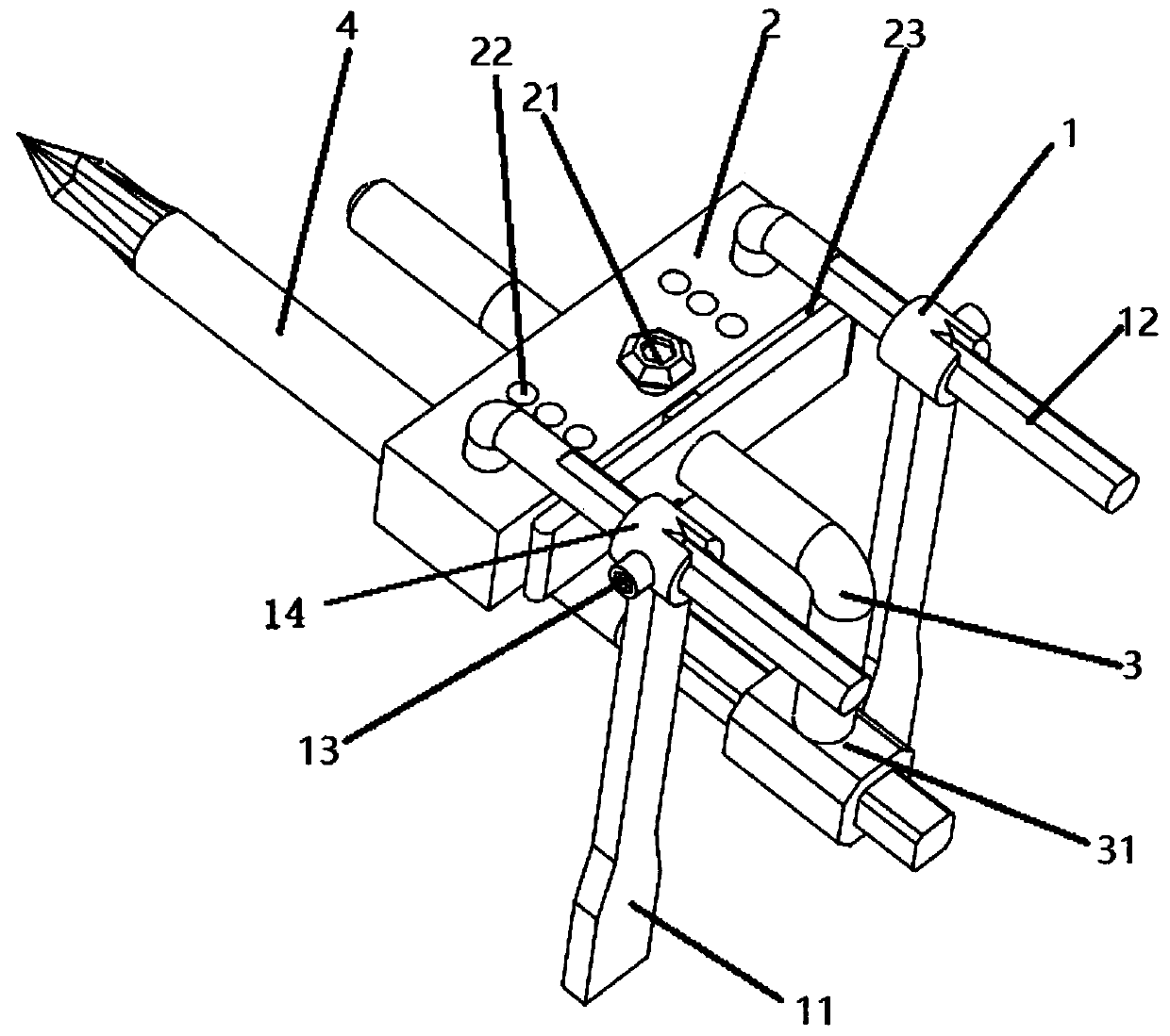
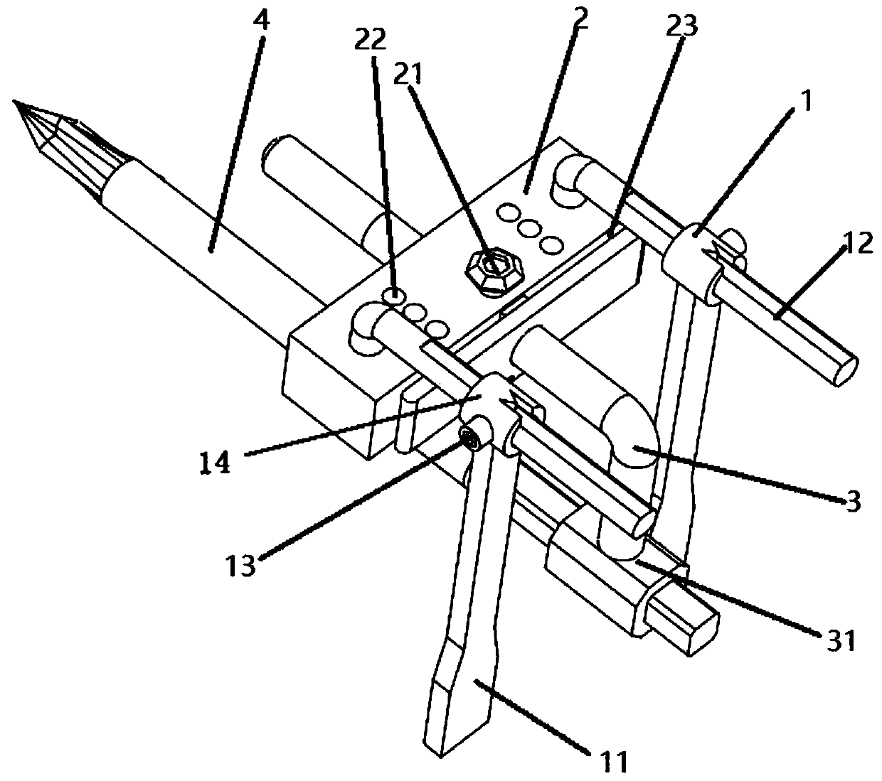
[0020] Such as figure 1 As shown, a distal femoral osteotomy device includes a femoral intramedullary fixation pin 4 fixed in the femoral medullary cavity, a connecting rod 3 installed on the femoral intramedullary fixation pin 4, a femoral section mounted on the connecting rod 3 The bone guide plate 2 and the osteotomy thickness measuring device 1 installed on the femoral osteotomy guide plate 2; the tip of the femoral intramedullary fixation needle 4 is provided with a "cross" groove for easy fixing in the femoral marr...


PUM


Abstract

The invention provides a distal femoral osteotomy device. The distal femoral osteotomy device comprises a femoral intramedullary fixation nail fixed in the femoral medullary cavity, a connecting rod mounted on the femoral intramedullary fixation nail, a femur osteotomy guide plate mounted on the connecting rod, and an osteotomy thickness gauge mounted on the femur osteotomy guide plate. The distalfemoral osteotomy device is convenient to adjust, ensures that the inner and outer osteotomy thickness is a predetermined osteotomy thickness and the osteotomy angles are the physiological varus andvalgus angles of a patient, achieves dynamic alignment in knee replacement, and is beneficial to rapid recovery of patients in order to improve the knee joint motion satisfaction of patients after replacement.

Description

technical field [0001] The invention relates to a distal femur osteotomy device. Background technique [0002] Total knee arthroplasty is a widely used and highly effective treatment for patients with advanced knee disease. Alignment technique is the basis of knee arthroplasty, which refers to the method of osteotomy and prosthesis installation according to the alignment during arthroplasty. The current traditional alignment method is mechanical axis alignment, that is, osteotomy and prosthesis installation are performed according to the mechanical axis of the lower limb formed by the center of the femoral head-knee joint center-ankle joint center. The rotation axis is perpendicular to the mechanical axis. For femoral osteotomy, the current practice is to find the anatomical axis of the femur by inserting the medullary canal rod through the medullary canal. With this anatomical axis as a reference, according to the degree of 5°, 6° or 7° ( This degree is obtained by measur...

Claims


Application Information

Patent Timeline
Patent Type & Authority Applications(China)
IPC IPC(8): A61B17/16A61B17/17
Inventor 谢杰苏伟平胡懿郃刘东昊卢玮刘田华王浩一
Owner XIANGYA HOSPITAL CENT SOUTH UNIV
Learn More