Drug-loaded fiber ring and preparation method and application thereof

A technology of annulus fibrosus and drug loading, which is applied in the field of cancer treatment to achieve the effect of inhibiting metastasis and invasion, high efficiency, and long-term stable and sustained release

Active Publication Date: 2019-06-14
DONGHUA UNIV +1
View PDF1 Cites 4 Cited by
  • Summary
  • Abstract
  • Description
  • Claims
  • Application Information

AI Technical Summary

Problems solved by technology

[0006] A search of domestic and foreign literature and patents on the preparation of drug-loaded annulus fiber shows that there is no relevant report on the preparation of drug-loaded annulus fibrosus by using electrospinning and homogenization technology to inhibit tumor metastasis and invasion.

Method used


Image

  • Drug-loaded fiber ring and preparation method and application thereof
  • Drug-loaded fiber ring and preparation method and application thereof
  • Drug-loaded fiber ring and preparation method and application thereof

Examples


Embodiment 1

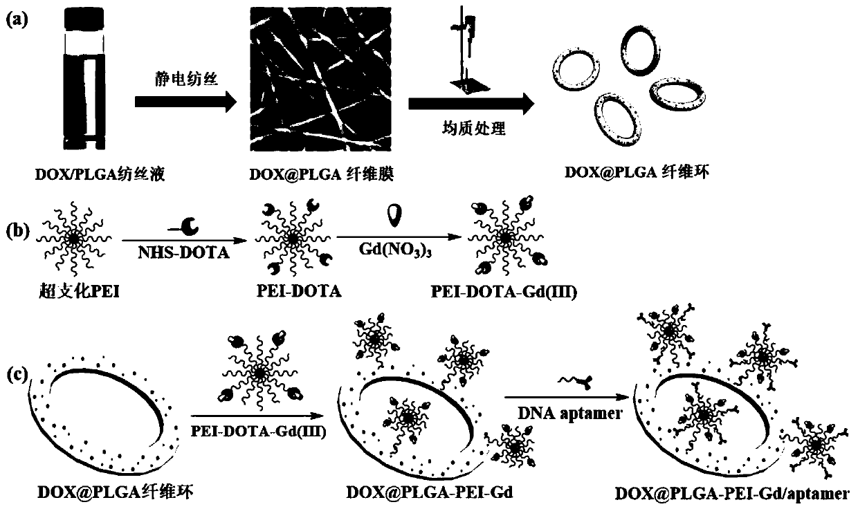
[0053] (1) Add 0.5g of polylactic acid-glycolic acid PLGA particles to 2mL of the mixed solution of N,N-dimethylformamide DMF and tetrahydrofuran THF, the volume ratio of DMF:THF is 1:3, magnetically stir for 4~ 8h, obtain PLGA spinning solution; Weigh 50mg doxorubicin hydrochloride DOX and join in PLGA spinning solution, stir vigorously for 48h, obtain DOX / PLGA spinning solution; Carry out electrospinning afterwards (spinning voltage 20kV, syringe pump flow rate is 0.6mL / h, the receiving distance is 15cm, the ambient temperature is 25°C, and the humidity is 30%), after vacuum drying for 48h, the DOX@PLGA nanofiber membrane loaded with anticancer drugs inside is obtained.

[0054] (2) Soak the DOX@PLGA nanofiber membrane obtained in step (1) in ultrapure water for 3 minutes, peel it off from the aluminum foil, and cut it into an area of ​​about 0.25 cm 2 The square fragments are then transferred to a polyvinyl alcohol PVA solution containing sodium chloride NaCl, wherein the c...

Embodiment 2

[0059] The present invention is characterized by scanning electron microscope SEM, ultraviolet-visible spectrophotometer UV-Vis, inductively coupled plasma spectrometer ICP, drug sustained release test, in vivo and in vitro magnetic resonance MR imaging performance test, tumor treatment and inhibition of metastasis and invasion evaluation. Various properties of the functionalized annulus fibrosus prepared in and its application potential in cancer therapy.

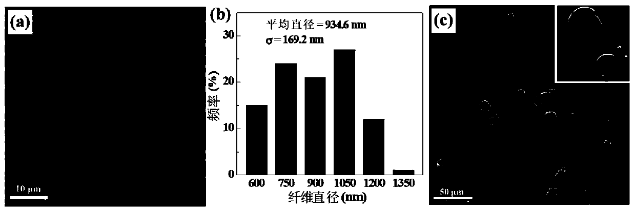
[0060] Scanning Electron Microscopy Test:

[0061] Characterize the morphology and size of the DOX@PLGA nanofibrous membrane obtained in the step (1) of Example 1 and the fiber ring of DOX@PLGA obtained in the step (2) of Example 1 by SEM, the results are as follows figure 2 As shown, it can be seen that the surface of DOX@PLGA nanofibers is smooth, the shape is uniform, and the fiber diameter is about 934.6nm; the average outer diameter of the DOX@PLGA fiber ring obtained after homogenization treatment is 11.1μm.
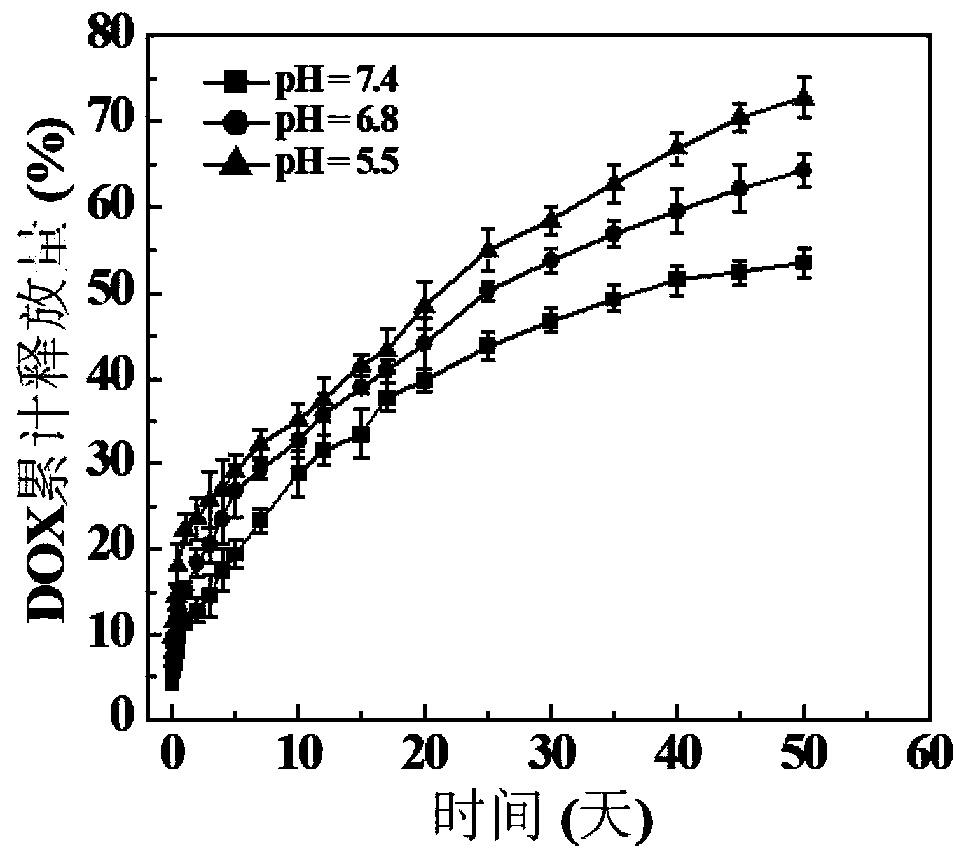
[006...

Embodiment 3

[0071] Hemolysis test:

[0072] The hemocompatibility of PLGA-PEI-Gd / aptamer in Comparative Example 1 was characterized by a hemolysis test. First, take 1 mL of fresh whole blood from a healthy person, centrifuge it, wash it with PBS three times, and discard the upper layer of plasma to obtain red blood cells (HRBCs). Dilute the obtained HRBCs 30 times with PBS solution, take out 0.2mL HRBCs dilution and add 0.8mL PBS as a negative control; take 0.2mL HRBCs dilution and add 0.8mL ultrapure water as a positive control. Dilute the HRBCs diluted 30 times above by 5 times with PBS, then weigh 2 mg, 4 mg, and 6 mg of PLGA-PEI-Gd / aptamer obtained in step (5) of Comparative Example 1, and add to 1 mL to dilute 5 times again In the final HRBCs suspension, 5 parallels were set for each concentration, and incubated in a constant temperature shaker at 37°C for 2h. Finally, centrifuge the control group and the HRBCs suspension containing the fibrosus material at a speed of 10000r / min fo...


PUM

PropertyMeasurementUnit
diameteraaaaaaaaaa
diameteraaaaaaaaaa
diameteraaaaaaaaaa

Abstract

The invention relates to a drug-loaded fiber ring and a preparation method and application thereof. The method comprises preparing a DOX/PLGA drug-loaded nanofiber membrane by electrospinning, and homogenizing to obtain the DOX/PLGA drug-loaded fiber ring. The surface is subjected to hyperbranched PEI chelating Gd(III) for MR imaging, and a modified DNA aptamer acts as a targeting molecule to be tightly bound to cancer cells, which can effectively inhibit tumor metastasis and invasion. The preparation method has the advantages of simple preparation process and good material biosafety. The ringmaterial is more likely to anchor the cancer cells and reduce the shedding of tumor cells. An internally loaded drug can effectively act on the tumor tissues for a long time, and effectively inhibitthe metastasis while killing the cancer cells. The fiber ring has broad application prospects in the field of cancer treatment.

Description

technical field [0001] The invention belongs to the field of cancer treatment, and in particular relates to a drug-loaded annulus fibrous and its preparation method and application. Background technique [0002] In recent years, cancer has become an important killer that endangers human life and health, and more than 90% of the deaths of cancer patients are caused by tumor metastasis and recurrence. Tumor cells have the characteristics of metastasis and invasion. Tumor cells detach from local tumor lesions in situ and enter the blood circulation system to become circulating tumor cells (CTCs). Once the tumor metastasizes to distant places, most treatments have limited curative effect and the prognosis is extremely poor. Once the tumor cells fall off into the blood, local treatment (surgery, radiotherapy) is ineffective. Even the systemic treatment - chemotherapy, has a certain killing effect on circulating tumor cells, but due to toxicity and other reasons, the intensity is...

Claims


Application Information

Patent Timeline
Patent Type & Authority Applications(China)
IPC IPC(8): A61K47/69A61K31/704A61K47/34A61K47/54A61K47/58A61K49/10A61K49/12A61P35/00A61P35/04
Inventor 史向阳刘勇肖云超涂文志
Owner DONGHUA UNIV
Why Eureka
Try Eureka