
Breast density quantitative calculation method in breast cancer risk assessment

A technology of risk assessment and breast density, applied in the field of medical image processing, can solve the problems of insufficient objective results, large influence, breast contour segmentation error, etc., achieve objective calculation results and reduce the burden of a large number of film readings

Active Publication Date: 2019-06-18
YANCHENG INST OF TECH
View PDF15 Cites 2 Cited by
  • Summary
  • Abstract
  • Description
  • Claims
  • Application Information

AI Technical Summary

Problems solved by technology

[0004] The semi-automatic gland segmentation method based on the manual interaction of radiologists is often greatly affected by the subjectivity of radiologists, and the results are not objective enough; although according to the guidance of Breast Imaging Reporting and Data System (BI-RADS), doctors’ images are also There are cases of fatigue reading, and visual fatigue often leads to errors in breast contour segmentation

Method used


Image

  • Breast density quantitative calculation method in breast cancer risk assessment
  • Breast density quantitative calculation method in breast cancer risk assessment
  • Breast density quantitative calculation method in breast cancer risk assessment

Examples


Embodiment 1

[0050] The quantitative calculation method of breast glandular tissue density according to one embodiment of the present invention specifically includes the following steps:

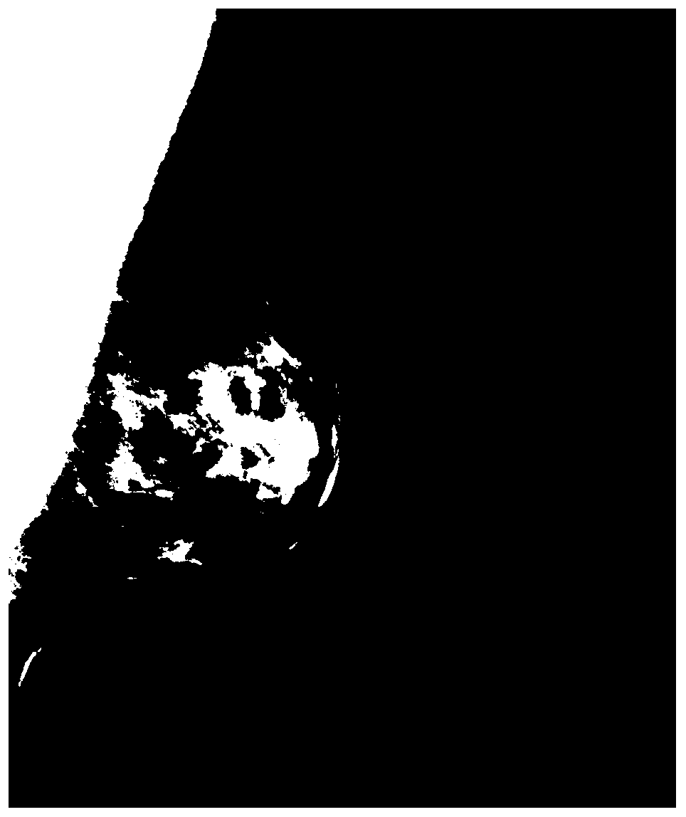
[0051] Step 1, for example figure 2 The mammography target MLO original image preprocessing shown in the figure, the specific operations include taking logarithm, inverting, and squaring the pixels to normalize and standardize the original less-standard images; and then using cubic spline interpolation to make the pixel size 2294*1914 The image is four times down-sampled to obtain an image with a pixel size of 574*479, and the processing speed of the subsequent algorithm is improved on the premise of ensuring that the useful information is not blurred.

[0052] Step 2. Segment a complete breast region from the preprocessed mammography MLO image, and the image includes three regions of pectoralis, breast and air background. First segment the outer contour of the breast, that is, the contour line of the ...


PUM


Abstract

The invention relates to a breast density quantitative calculation method in breast cancer risk assessment. The method specifically comprises the following steps that an original image of a breast molybdenum target is preprocessed, and histogram equalization, Gaussian filtering and down-sampling are carried out; a breast area is segmented from the original image of the breast molybdenum target, and the outer edge line of the breast and the inner edge line of the breast muscle are detected; fuzzy C-means are used for carrying out unsupervised clustering on pixels in the breast area; features ofa cluster region are extracted, obtained clusters are merged and classified into glands and fat, and the clusters are subjected to dichotomy recognition; dichotomy is carried out on the breast area,and a linear discriminant LDA classifier is trained; the size of the glands in the breast area is calculated according to a classification result. According to the breast density quantitative calculation method, full-automatic segmentation of breast tissue in the breast can be realized, and the burden of an imaging doctor is greatly reduced while an objective result is given.

Description

technical field [0001] The invention relates to the field of medical image processing, in particular to a method for quantitative calculation of breast density in breast cancer risk assessment. Background technique [0002] There are two types of breast cancer risk assessment models, one is the empirical risk model such as the Gail model, and the other is the IBIS model that focuses on gene mutations. Among them, the Gail model is the most commonly used. This model is used to evaluate and calculate the risk value of individualized breast cancer, including multiple risk factors related to breast cancer. The model gives different weight coefficients to each risk factor, and a quantitative weighted combination is calculated. cancer risk value. The risk factors in the Gail model include weight, age, breast gland type, menarche and menopause time, breastfeeding status, BMI index, smoking or drinking, mental stress, etc., and the breast gland type mainly depends on experienced ex...

Claims


Application Information

Patent Timeline
IPC IPC(8): A61B5/00
Inventor 王东洋王银杰陈永明
Owner YANCHENG INST OF TECH
Learn More