Capsule type endoscope and control method thereof

A technology of capsule endoscope and control method, which is applied in the field of medical equipment, can solve the problem that the micro-morphological structure cannot be displayed by photographing, and achieve the effect of improving the diagnosis rate of lesions

Pending Publication Date: 2019-07-12
安翰科技(武汉)股份有限公司
View PDF12 Cites 6 Cited by
  • Summary
  • Abstract
  • Description
  • Claims
  • Application Information

AI Technical Summary

Problems solved by technology

[0005] However, the existing capsule endoscopes only display the epidermal images inside the digestive organs of the subject for subsequent diagnosis, and often many early lesions appear on the surface of mucosal cells. The normal lens of the speculum cannot capture the displayed

Method used


Image

  • Capsule type endoscope and control method thereof
  • Capsule type endoscope and control method thereof
  • Capsule type endoscope and control method thereof

Examples


Embodiment Construction

[0033] The present invention will be described in detail below with reference to the specific embodiments shown in the accompanying drawings. However, these embodiments do not limit the present invention, and structural, method, or functional changes made by those skilled in the art according to these embodiments are all included in the protection scope of the present invention.

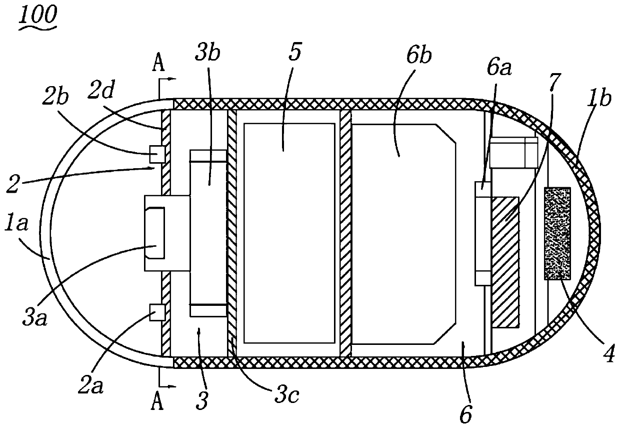
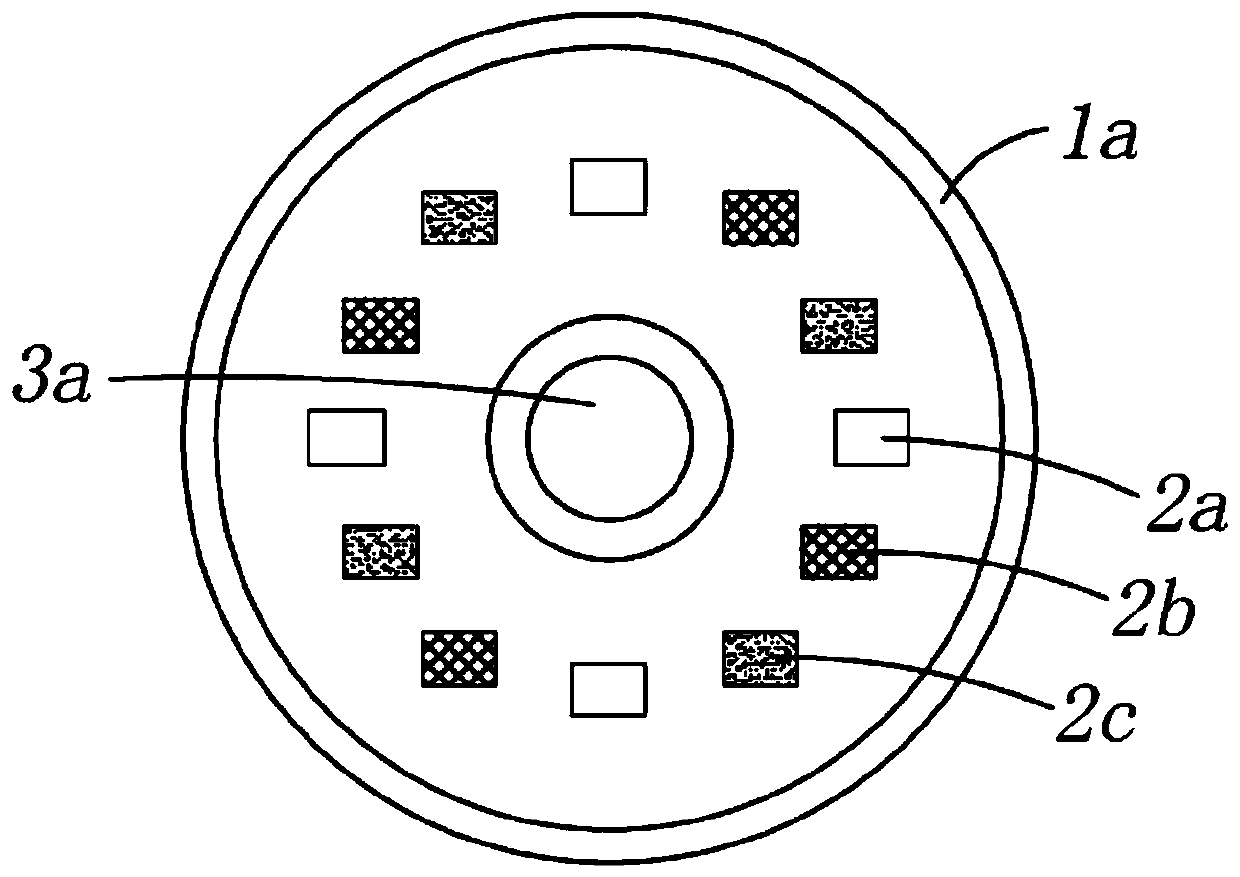
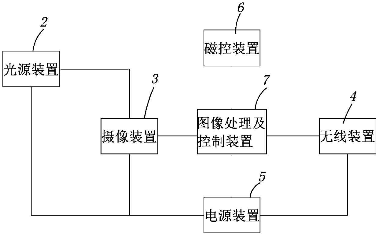
[0034] refer to Figure 1 to Figure 3 As shown, in a preferred embodiment of the present invention, the capsule endoscope is used to introduce into the interior of the subject and capture an internal image of the subject, and the capsule endoscope 100 includes a housing and a light source accommodated in the housing. Device 2 , photographing device 3 , wireless device 4 , power supply device 5 , magnetron device 6 , and image processing and control device 7 . The casing includes a main casing 1b and an optical dome 1a enclosed at one end of the main casing 1b, the optical dome 1a is located at the f...


PUM


Abstract

The invention provides a capsule type endoscope, which comprises a shell, a shooting device and a light source device, wherein the shooting device and the light source device are accommodated in the shell; the light source device irradiates illumination light on the inner surface of a subject; the shooting device shoots an internal image of the subject based on the illumination light; the shell comprises a main shell and an optical dome packaged at one end of the main shell; the optical dome is positioned at the front ends of the shooting device and the light source device; the light source device comprises a first light source module and a second light source module; the wavelength ranges of light emitted by the first light source module and the second light source module are different; the first light source module and the second light source module are arranged at intervals around the shooting device. According to the capsule type endoscope, by arranging at least two different lightsource devices, common focus images can be normally and clearly displayed, and forms of different layers of mucosa images can be obtained, so that deep focus is found, and the focus diagnosis rate iseffectively improved.

Description

technical field [0001] The invention relates to the technical field of medical devices, in particular to a capsule endoscope and a control method thereof. Background technique [0002] In recent years, in the field of endoscopy, a swallowable capsule endoscope provided with an imaging function and a wireless communication function has been proposed. Capsule-type endoscopes are ingested from the mouth of the subject for observation (inspection) until they are naturally excreted from the subject. While the inside thereof (ie, the inside of the digestive tract) is moved, images of the inside of the digestive tract of the subject are sequentially captured at intervals of, for example, 0.5 seconds. [0003] In addition, while the capsule endoscope is moving in the alimentary canal of the subject, images captured by the capsule endoscope are sequentially transmitted from the capsule endoscope to an external receiving device by wireless communication. The receiving device has a f...

Claims


Application Information

Patent Timeline
Patent Type & Authority Applications(China)
IPC IPC(8): A61B1/04A61B1/06A61B5/07
CPCA61B1/041A61B1/0638A61B1/0661A61B5/07A61B1/00158A61B1/00016A61B1/0607A61B1/063A61B1/000094A61B1/0655A61B1/00006A61B1/00195A61B5/0084A61B5/0086
Inventor 明繁华彭航宇王蓉丰波周念琪
Owner 安翰科技(武汉)股份有限公司
Why Eureka
Try Eureka