Lung tumor segmentation method for large-area adhesion of lung boundary tissue in CT image

A CT image, large-area technology, applied in the field of lung tumor segmentation, can solve the problem of poor segmentation accuracy of large adhesion-type lung tumors

Active Publication Date: 2019-07-19
HEBEI UNIVERSITY
View PDF10 Cites 4 Cited by
  • Summary
  • Abstract
  • Description
  • Claims
  • Application Information

AI Technical Summary

Problems solved by technology

[0005] The purpose of the present invention is to provide a method for segmenting lung tumors with a large area of ​​adherent pulmonary border tissue in CT images, so as to solve the problem of poor segmentation accuracy of existing methods for large adherent lung tumors

Method used


Image

  • Lung tumor segmentation method for large-area adhesion of lung boundary tissue in CT image
  • Lung tumor segmentation method for large-area adhesion of lung boundary tissue in CT image
  • Lung tumor segmentation method for large-area adhesion of lung boundary tissue in CT image

Examples


Embodiment Construction

[0051] The specific implementation process of the present invention adopts the following computer software and hardware conditions to realize, but is not limited to the following conditions: Lenovo desktop computer, CPU is Pentium Dual-Core CPU E5800@3.20GHz, graphics card is NVIDIA GeForce GT 430GPU, memory 4GB, operation The system is Window 7, and the software programming language uses Matlab 2009.

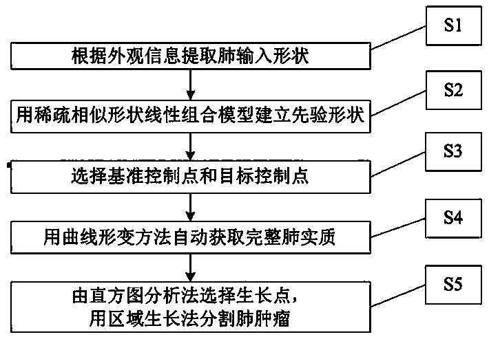
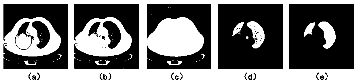
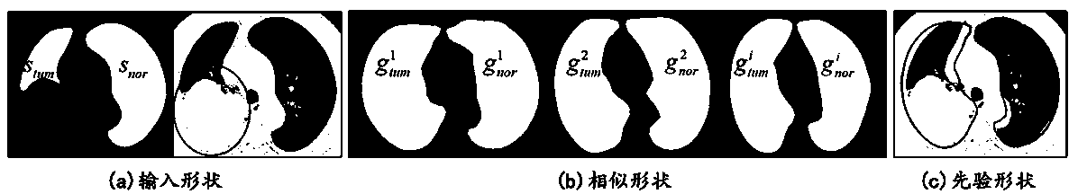
[0052] The basic process of the lung tumor segmentation method with large-area adhesion of lung border tissue in CT images of the present invention is as follows: figure 1 As shown in the figure, first, according to the image appearance information, the lung parenchyma with large continuity error is segmented by Otsu threshold and morphological opening and closing operation method, and this is used as the input shape. Shape priors are then constructed using a sparse similar shape linear combination model. Then select the deformation curve and its control points on the prior sh...


PUM


Abstract

The invention discloses a lung tumor segmentation method for large-area adhesion of lung boundary tissue in a CT image. The method comprises the following steps: extracting left and right pulmonary lobe parenchyma shapes from a lung CT image containing a large tumor, and constructing an input shape; then, according to the tumor-free lung CT image, constructing a prior shape by using a sparse similar shape linear combination model; selecting a deformation curve and a control point thereof on a priori shape, and selecting a target curve and a control point thereof on an input shape; using a curvilinear transformation method for correcting a large continuous error on the pulmonary parenchyma shape (namely an input shape), so that a complete pulmonary parenchyma contour including tumors is obtained, and a pulmonary parenchyma image is further obtained; and finally, segmenting the lung tumor on the lung parenchyma image by using a region growing method.

Description

technical field [0001] The invention relates to a method for processing CT images, in particular to a method for segmenting lung tumors with large-area adhesion of lung boundary tissue in CT images. Background technique [0002] Among all clinical imaging modalities, computed tomography (CT) is a direct and effective modality for the extraction of lung and its lesion characteristics, disease diagnosis and therapeutic efficacy assessment. Accurate segmentation of lung tumors is crucial for precise radiation therapy planning and treatment response evaluation, and is a research hotspot in the imaging diagnosis of lung cancer. However, the segmentation of large tumors connected to or invading anatomical structures such as the chest wall, thoracic spine, diaphragm, mediastinum, or heart is still a topic that requires in-depth study. [0003] Large tumors have a large area of ​​adhesion to surrounding tissues such as the chest wall and mediastinum, which seriously damages the lun...

Claims


Application Information

Patent Timeline
Patent Type & Authority Applications(China)
IPC IPC(8): G06T7/00G06T7/11G06T7/136
CPCG06T7/0012G06T2207/10081G06T2207/30061G06T2207/30096G06T7/11G06T7/136
Inventor 张欣王洁王兵
Owner HEBEI UNIVERSITY
Why Eureka
Try Eureka