Implantable observation device for animal liver living body imaging

A technology of in vivo imaging and animal liver, which is applied in the field of medical devices to achieve the effect of maintaining long-term stability, avoiding abdominal exposure surgery, and maintaining exposure

Inactive Publication Date: 2019-07-23
FOURTH MILITARY MEDICAL UNIVERSITY
View PDF4 Cites 4 Cited by
  • Summary
  • Abstract
  • Description
  • Claims
  • Application Information

AI Technical Summary

Problems solved by technology

[0004] The purpose of the present invention is to provide an implantable observation device for living animal liver imaging, which solves the problem in the prior art that requires repeated exposure operations to dynamically track the liver observation area

Method used


Image

  • Implantable observation device for animal liver living body imaging
  • Implantable observation device for animal liver living body imaging
  • Implantable observation device for animal liver living body imaging

Examples


Embodiment Construction

[0017] The present invention will be described in detail below in conjunction with the accompanying drawings and specific embodiments.

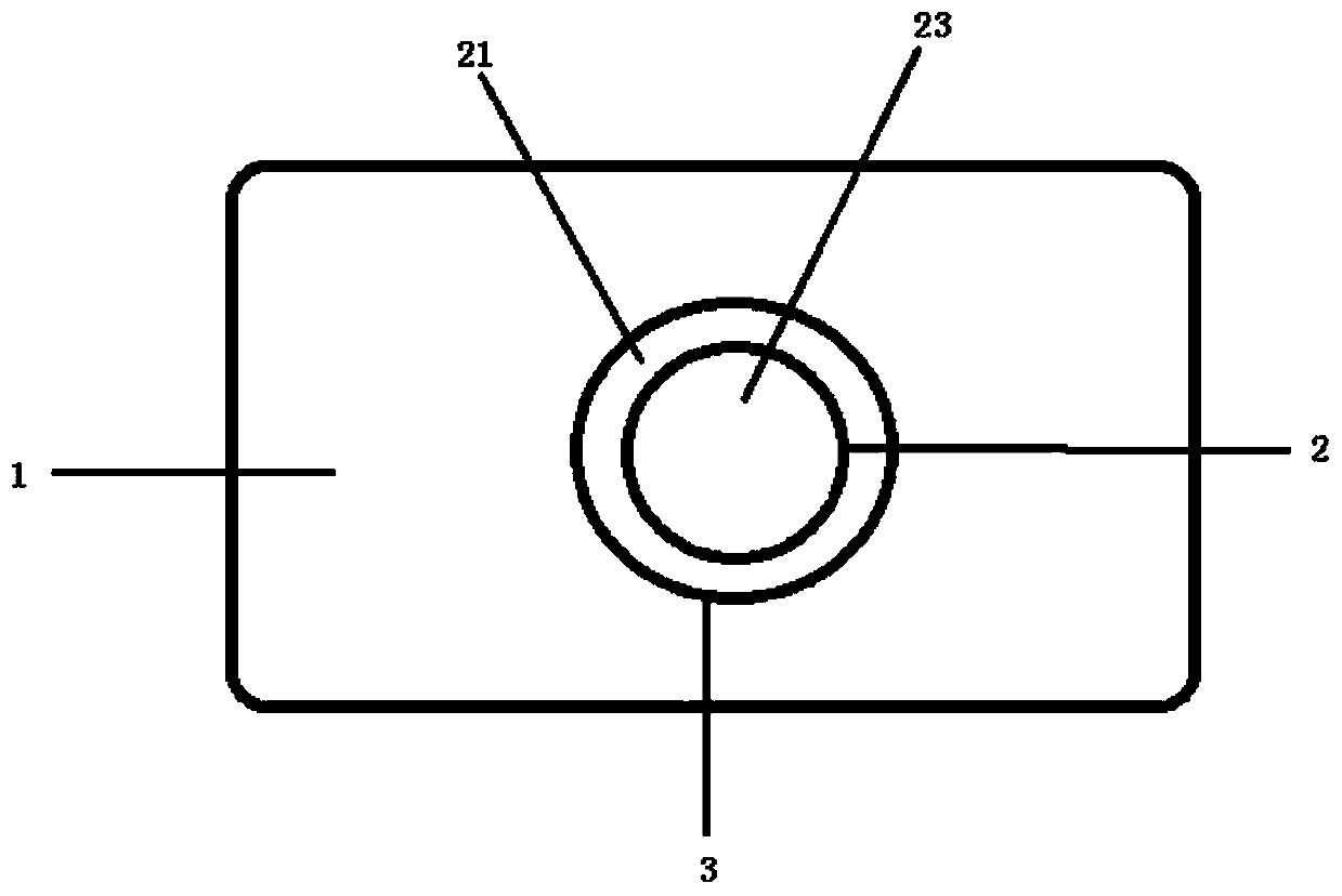
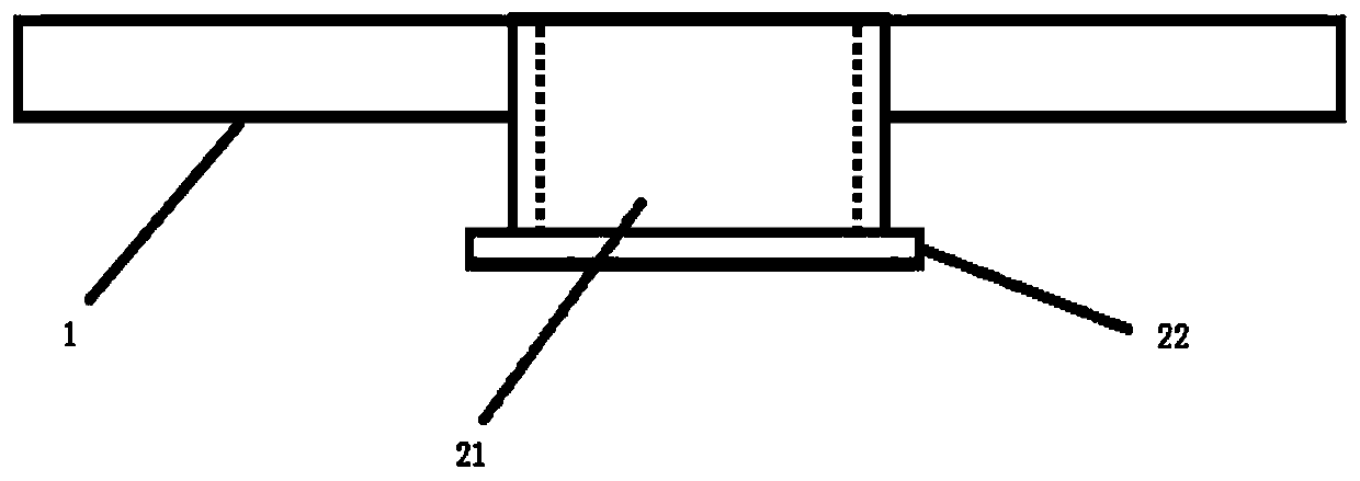
[0018] An implantable observation device for animal liver living imaging of the present invention, such as figure 1 As shown, including object plate 1 and observation block 2; as figure 2 As shown, the observation block 2 includes a ring 21, one end of the ring 21 is provided with a boss 22, and the other end of the ring 21 is provided with a cover glass 23; The hole 3 , the through hole 3 is arranged in the center of the loading plate 1 . The circular ring 21 is clearance fit with the carrier plate 1 . One end of the ring 21 provided with a cover glass 23 is provided with a groove concentric with the ring 21, and the cover glass 23 is arranged in the groove. The cover glass 23 is provided with a positioning scale grid.

[0019] The object loading plate 1 is a rectangular plate whose size matches the microscope object loading platform. ...


PUM


Abstract

The present invention discloses an implantable observation device for animal liver living body imaging. The implantable observation device comprises a carrier plate, a through-hole is arranged in thecarrier plate, and an observation block is arranged in the through-hole; the observation block comprises a round ring, an outer wall of one end of the round ring is fixedly connected with a boss in acircumferential direction, and the other end of the round ring is fixedly provided with a cover glass; and the round ring is matched with the through-hole and one end of the round ring fixed connectedwith the boss protrudes from the through-hole. When the implantable observation device is used, liver tissues are pressed to contact with the cover glass and then the liver tissues are adhered to a sphere through biological glue; then animal abdominal skin and muscles are sutured and fixed on the boss by a pouch to complete an implantation process of the observation block. When animal liver needsto be observed, the observation block is placed in the through-hole of the carrier plate and placed on a confocal microscope to perform imaging observation. After the observation, the observation block is separated from the carrier plate. The implantable observation device avoids repeated abdominal exposure surgeries, maintains long-term stable exposure of the local liver tissues, and is beneficial for the repeated long-term observation.

Description

technical field [0001] The invention belongs to the technical field of medical devices, and relates to an implantable observation device for living animal liver imaging. Background technique [0002] In liver research experiments, it is an important research method to use single-photon or two-photon confocal microscopes to dynamically observe animal livers. The traditional in vivo imaging method of animal liver is to cut the abdominal skin and muscle to expose the entire liver surface, place it on a glass slide, and directly use a confocal microscope to perform dynamic imaging of the exposed animal liver surface in vivo. After the imaging was completed, the liver was returned, and the muscles and skin of the abdomen were sutured layer by layer. The next time the skin and muscles were cut, the original liver was exposed and repeated. [0003] Although this method can be used to observe the physiological and pathological changes of the liver operation, it cannot be repeated m...

Claims


Application Information

Patent Timeline
Patent Type & Authority Applications(China)
IPC IPC(8): A61B5/00
CPCA61B5/6847A61B5/6867A61B2503/40
Inventor 樊俊俊李晓祥毕龙袁志
Owner FOURTH MILITARY MEDICAL UNIVERSITY
Why Eureka
Try Eureka