Automatic liver tumor classification method and device based on multi-stage CT image analysis

A CT image and automatic classification technology, applied in image analysis, neural learning methods, image enhancement, etc., can solve the problems of improving recognition rate, unclear display of lesion sites, limited tumor characteristic information, etc., and achieve high precision results

Pending Publication Date: 2020-03-27
BEIJING INSTITUTE OF TECHNOLOGYGY
View PDF6 Cites 16 Cited by
  • Summary
  • Abstract
  • Description
  • Claims
  • Application Information

AI Technical Summary

Problems solved by technology

[0005] 1. The size, shape, and location of tumors in different patients are different, and the lesion site is not displayed clearly, which brings challenges to traditional identification methods
[0006] 2. The recognition method based on traditional machine learning needs to manually design feature extraction methods for different types of liver cancer. The quality of the method design directly affects the final recognition performance
[0007] 3. Most of the existing methods are trained based on CT images in the plain scan period. Such data can reflect very limited tumor characteristic information, and the multi-phase contrast-enhanced data has not been comprehensively used to improve the recognition rate.

Method used


Image

  • Automatic liver tumor classification method and device based on multi-stage CT image analysis
  • Automatic liver tumor classification method and device based on multi-stage CT image analysis

Examples


Embodiment Construction

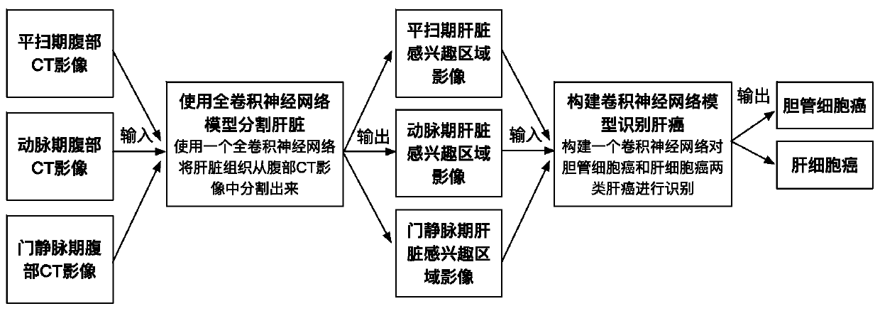
[0021] Such as figure 1 As shown, this method for automatic classification of liver tumors based on multi-phase CT image analysis includes the following steps:

[0022] (1) Acquire contrast-enhanced abdominal CT scan images of patients with cholangiocarcinoma and hepatocellular carcinoma, save them as arterial phase, portal venous phase, and delayed phase, and diagnose the type of liver cancer to which all data belong, as The gold standard for model training;

[0023] (2) Construct a three-dimensional fully convolutional neural network segmentation model, use the image data of cholangiocarcinoma and hepatocellular carcinoma collected in step (1) as the input of the model for learning, and learn the intrinsic characteristics of liver tissue at each stage through the model Fully automatic training and learning, so as to segment it from abdominal CT images, as the region of interest of the subsequent liver cancer recognition model;

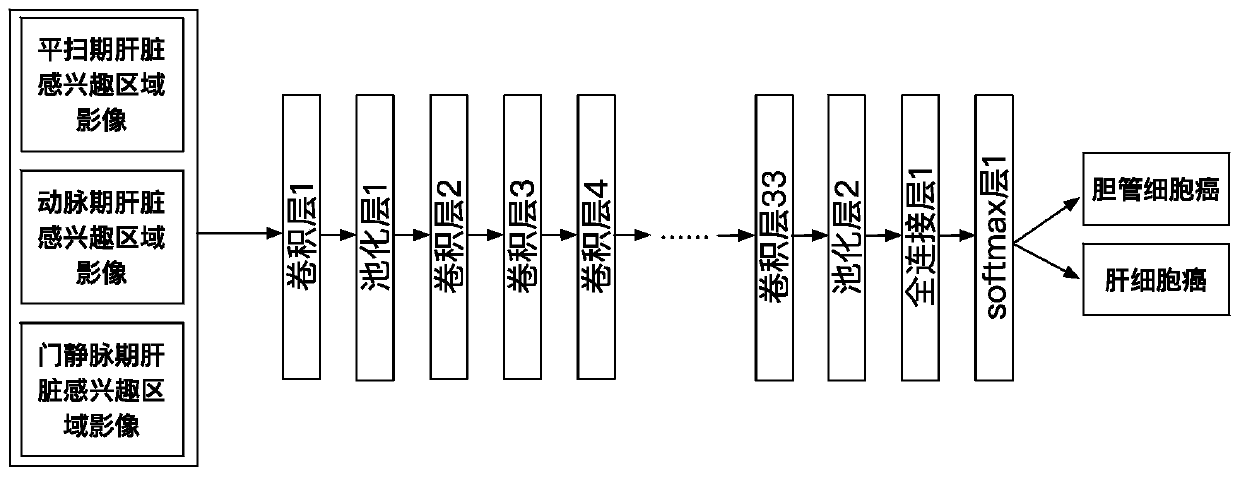
[0024] (3) Construct a three-dimensional con...


PUM


Abstract

According to the liver tumor automatic classification method and device based on multi-stage CT image analysis, full-automatic bile duct cell carcinoma and hepatocellular carcinoma can be recognized,and a high-precision bile duct cell carcinoma and hepatocellular carcinoma recognition model is obtained. The method comprises the following steps: (1) acquiring a contrast-enhanced abdominal CT scanning image, storing the contrast-enhanced abdominal CT scanning image as an arterial phase, a portal vein phase and a delay phase, and carrying out definite diagnosis on liver cancer categories to which all data belong to serve as a model training gold standard; (2) constructing a three-dimensional full convolutional neural network segmentation model, and segmenting the intrinsic characteristics ofthe liver tissue in each stage from the abdominal CT image through model training learning; (3) constructing a three-dimensional convolutional neural network classification model; and inputting the image data obtained by segmentation into a classification model for training, so as to enable the model to perform joint learning and training on the cancer features in multiple periods, thereby predicting the category to which the cancer belongs, comparing the prediction result with a gold standard, and supervising the training process of the model in a loss value feedback mode.

Description

technical field [0001] The present invention relates to the technical field of medical image processing, in particular to a method for automatic classification of liver tumors based on multi-phase CT image analysis, and an automatic classification device for liver tumors based on multi-phase CT image analysis, which is mainly applicable to the identification of liver cancer based on CT images Research areas. Background technique [0002] In recent years, the incidence of cholangiocarcinoma is on the rise. It is located in the liver and has no obvious clinical symptoms in the early stage. It is easy to be misdiagnosed as hepatocellular carcinoma. However, its treatment is completely different from that of hepatocellular carcinoma. The best timing of surgery has been lost during the disease, therefore, the early diagnosis of cholangiocarcinoma has important clinical significance. The traditional diagnosis of cholangiocarcinoma is mainly judged by doctors observing CT images o...

Claims


Application Information

Patent Timeline
Patent Type & Authority Applications(China)
IPC IPC(8): G06K9/62G06K9/32G06N3/04G06N3/08G06T7/11
CPCG06T7/11G06N3/08G06T2207/10081G06V10/25G06N3/045G06F18/241G06F18/253
Inventor 宋红陈磊杨健范敬凡
Owner BEIJING INSTITUTE OF TECHNOLOGYGY
Why Eureka
Try Eureka