Excision side generating method and device of liver operation

A technology for generating devices and cutting surfaces, applied in the field of computers, can solve the problems of not considering the difficulty and feasibility of surgery, and not providing lesions, and achieve the effects of reducing the possibility of postoperative complications, improving the ratio of residual liver, and reducing difficulty.

Active Publication Date: 2018-07-20
浙江京新术派医疗科技有限公司
View PDF3 Cites 15 Cited by
  • Summary
  • Abstract
  • Description
  • Claims
  • Application Information

AI Technical Summary

Problems solved by technology

However, at present, when generating the surgical cutting surface, the relationship between the lesion and the blood supply and blood return area is not

Method used


Image

  • Excision side generating method and device of liver operation
  • Excision side generating method and device of liver operation
  • Excision side generating method and device of liver operation

Examples


Embodiment 1

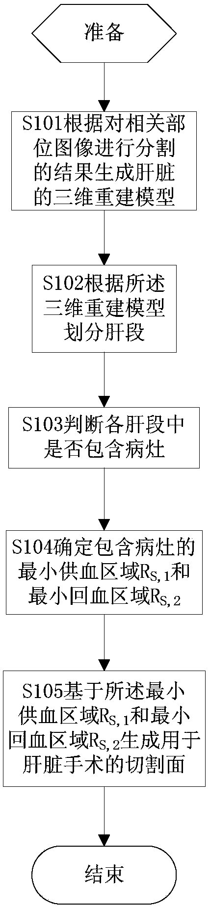
[0048] like figure 1 As shown, in the first embodiment of the present invention, a three-dimensional reconstruction model is generated according to the CT image segmentation results of the relevant parts before surgery. The blood return area generates a surgical cut surface of the liver for removal of the lesion.

[0049] S101. Generate a three-dimensional reconstruction model of the liver according to the image segmentation result.

[0050] First, CT or MRI image acquisition of relevant parts is performed. CT acquisition adopts liver CT multi-phase enhanced scanning technology. Taking advantage of the difference in the time when the developer enters different parts, images are collected in different periods to obtain enhanced images for different targets. Usually, the image of the liver is divided into five phase phases, namely the liver phase, which is used for liver segmentation; the lesion phase, which is an enhanced image of the lesion, which is used for lesion segment...

Embodiment 2

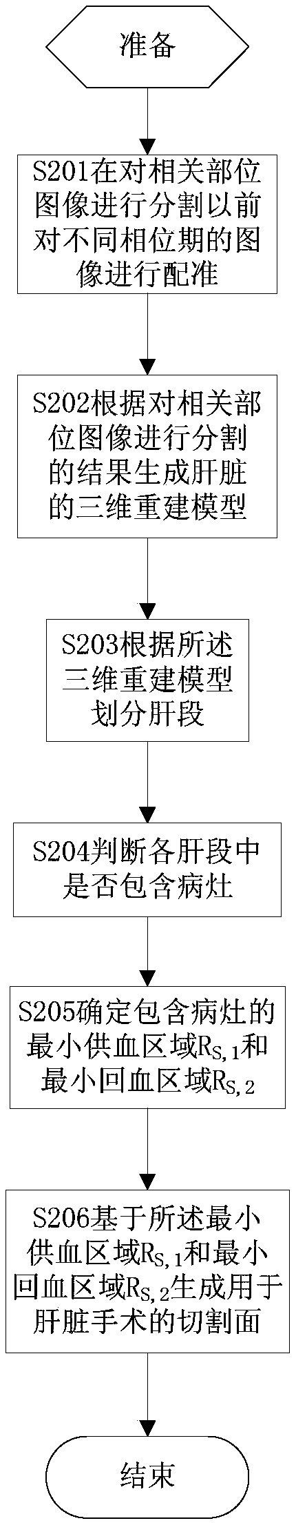
[0063] like figure 2 As shown, in the second embodiment of the present invention, when reconstructing a three-dimensional model, images in different phase periods are registered to ensure the accuracy of the reconstruction results; In the segment, the surgical cut plane of the liver is generated according to the minimum blood supply area and the minimum blood return area.

[0064] S201 , registering images in different phase periods.

[0065] Contrast agents are used when acquiring CT or MRI images of the liver, and images are acquired for different relevant parts in different periods to obtain images that are enhanced for different targets. Due to the displacement of the patient, the collected images have errors, so it is necessary to perform denoising through registration, so as to minimize the displacement error between images of different phase periods during 3D reconstruction and ensure the accuracy of the reconstruction results.

[0066] Taking the registration of liv...

Embodiment 3

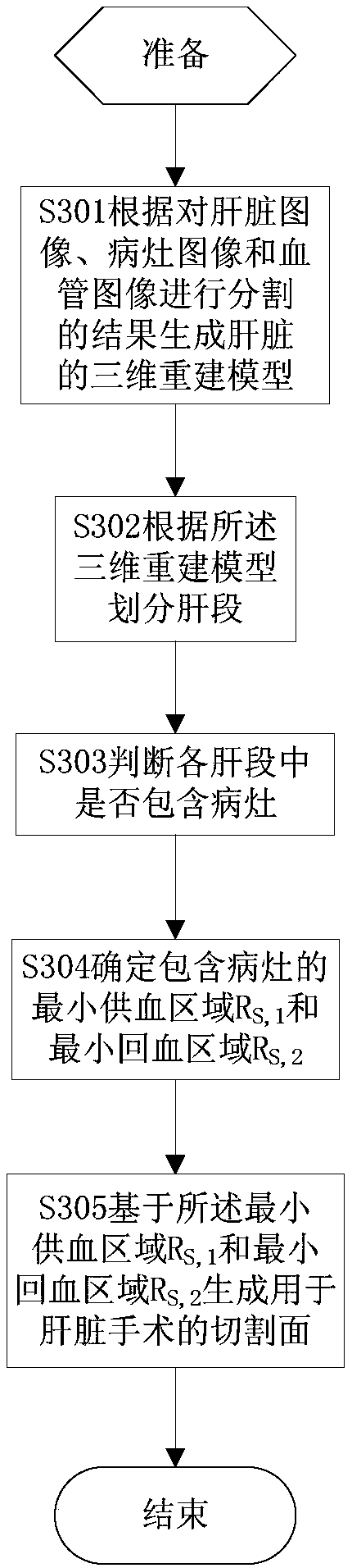
[0078] like Figure 3a As shown, in the third embodiment of the present invention, a three-dimensional reconstruction model of the liver is generated according to the results of segmenting the images of the liver, lesions and blood vessels, and after the liver segment is divided, the minimum blood supply area of ​​the liver segment including the lesion is determined and minimal blood return area, and generate a surgical cut surface of the liver.

[0079] S301. Generate a three-dimensional reconstruction model of the liver according to the result of segmenting the liver, the lesions and the blood vessels.

[0080] Taking the preoperative CT or MRI images of the relevant parts of the patient as input, the liver is automatically segmented to obtain preliminary segmentation results. On the basis of this result, the user can manually make local adjustments to the segmentation results. CT or MRI images are composed of multiple consecutive slice images, and the user only needs to m...


PUM


Abstract

The invention discloses an excision side generating method and device of a liver operation. The method comprises that a 3D reconstruction model is generated according to an image segmentation result;liver segments are divided according to the 3D reconstruction model; whether each liver segment includes a focus is determined; a minimal blood supply area RS,1 and a minimal blood return area RS,2 including the focus are determined; and an excision side of the liver operation is generated on the basis of the RS,1 and RS,2. Registering is carried out; and the 2D reconstruction model is generated according to a liver image, a focus image and a blood vessel image segmentation result. When the focus only exists in one liver segment, the excision side of the liver operation is generated by combining the minimal blood supply area with the minimal blood return area of the liver segment with the focus; and when multiple liver segments include focuses, the excision side of the liver operation is generated according to an area by combining the minimal blood supply areas the minimal blood areas of the liver segments and an excision area. Thus, a doctor is helped make a better operation plan, andmore healthy liver tissues can be reserved.

Description

technical field [0001] The invention relates to computer technology, in particular to computer-aided preoperative planning technology. Background technique [0002] Currently, partial hepatectomy is the first choice for the treatment of liver cancer and malignant tumors. Traditional partial hepatectomy surgery generally adopts the whole or multi-segment resection method based on the anatomy of Cubit's liver segment. [0003] In the traditional surgical planning method, the boundary of surgical resection mainly depends on the subjective judgment of the clinician and largely depends on the clinical experience of the doctor. For the variation of blood vessel structure and distribution, it is difficult to judge by direct observation of CT or MRI images. Wrong judgment will cause blood vessels to be cut by mistake during surgery, which will endanger the life safety of patients. Due to the lack of accurate preoperative planning, healthy liver tissue may be removed by mistake, af...

Claims


Application Information

Patent Timeline
IPC IPC(8): G06T7/11G06T7/12G06T7/187G06T7/30G06T7/00G06T17/00G06F19/00
CPCG06T7/0012G06T17/00G06T2207/30056G06T2207/30096G06T2207/30101G06T2210/41G06T2207/10088G06T2207/10081
Inventor 谢卫国王磊徐宁周龙宋晓堃
Owner 浙江京新术派医疗科技有限公司
Why Eureka
Try Eureka