Image segmentation method based on feature extraction and denoising
A feature extraction and image segmentation technology, applied in the field of image processing, can solve the problems of inaccurate segmentation, interference with gestational sac segmentation, and the inability of algorithms to be independently competent.
- Summary
- Abstract
- Description
- Claims
- Application Information
AI Technical Summary
Problems solved by technology
Method used
Image
Examples
Embodiment 1
[0088] An image segmentation method based on feature extraction and denoising, including the gestational sac segmentation process and the yolk sac-germ development state segmentation process;
[0089] The gestational sac segmentation process includes the following steps:
[0090] Step 1: Input B-ultrasound images such as figure 1 (a) and figure 2 (a);
[0091] Step 2: Obtain the binary image of the B-ultrasound image through image segmentation and feature extraction;
[0092] Step 3: Select the area where the gestational sac is located to obtain the binary image of the gestational sac, such as figure 1 (b) and figure 2 (b), the processing steps are as follows:
[0093] 3.1) Use the mask method to select the area where the gestational sac is located to obtain an image;
[0094] 3.2) According to the image binary image obtained in 3.1, remove the edge noise to obtain the edge noise-removed image.
[0095] Step 4: Image feature extraction, the processing steps are as fol...
Embodiment 2
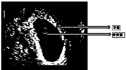
[0143] Such as figure 1 Shown is the division of the gestational sac and yolk sac
[0144] (1) Image description;
[0145] (2) Read in an original B-ultrasound grayscale image src_before, as attached figure 1 (a);
[0146] (3) Use the level set algorithm to segment the image of src_before, and then select the area where the gestational sac is located according to the segmented B-ultrasound image (in order to remove the effusion, dark areas, etc., and the grayscale features are similar to the gestational sac part to segment the gestational sac The impact of the results), and then perform feature extraction and denoising operations by observing the characteristics of the gestational sac, and finally obtain the binary image GS_img of the gestational sac, as shown in the attached figure 1 (b);
[0147] (4) Grayscale the binary image GS_img of the gestational sac to obtain the binary result image GS_Gray of the gestational sac and output it, as attached figure 1 (c);
[0148]...
Embodiment 3
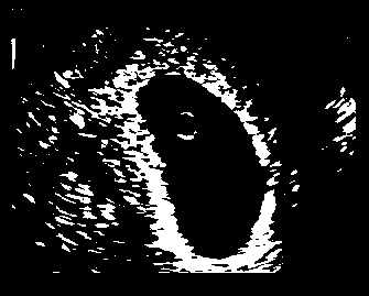
[0154] Such as figure 2 Shown is the division of the gestational sac and germ
[0155] (1) Image description;
[0156] (2) Read in an original B-ultrasound grayscale image src_before, as attached figure 2 (a);
[0157] (3) Use the level set algorithm to segment the image of src_before, and then select the area where the gestational sac is located according to the segmented B-ultrasound image (in order to remove the effusion, dark areas, etc., and the grayscale features are similar to the gestational sac part to segment the gestational sac The impact of the results), and then perform feature extraction and denoising operations by observing the characteristics of the gestational sac, and finally obtain the binary image GS_img of the gestational sac, as shown in the attached figure 2 (b);
[0158] (4) Grayscale the binary image GS_img of the gestational sac to obtain the binary result image GS_Gray of the gestational sac and output it, as attached figure 2 (c);
[0159]...
PUM
 Abstract
Description
Claims
Application Information
 - R&D
- Intellectual Property
- Life Sciences
- Materials
- Tech Scout
- Unparalleled Data Quality
- Higher Quality Content
- 60% Fewer Hallucinations
Browse by: Latest US Patents, China's latest patents, Technical Efficacy Thesaurus, Application Domain, Technology Topic, Popular Technical Reports.
© 2025 PatSnap. All rights reserved.Legal|Privacy policy|Modern Slavery Act Transparency Statement|Sitemap|About US| Contact US: help@patsnap.com


