Liver three-dimensional CT image lesion area automatic extraction system and method

A technology of CT images and lesion areas, applied in the field of liver three-dimensional image processing methods and systems, can solve problems such as misjudgment, single angle of lesion area feature selection, and easy to identify loopholes in patient areas, so as to improve effectiveness and reliability performance, ensuring precision and accuracy

Pending Publication Date: 2019-08-16
THE AFFILIATED HOSPITAL OF QINGDAO UNIV
View PDF1 Cites 1 Cited by
  • Summary
  • Abstract
  • Description
  • Claims
  • Application Information

AI Technical Summary

Problems solved by technology

However, the random forest algorithm-based liver CT image lesion recognition method of the patent application only uses the image grayscale te

Method used


Image

  • Liver three-dimensional CT image lesion area automatic extraction system and method
  • Liver three-dimensional CT image lesion area automatic extraction system and method
  • Liver three-dimensional CT image lesion area automatic extraction system and method

Examples


Embodiment Construction

[0025] Embodiments of the present invention are described in detail below, examples of which are shown in the drawings, wherein the same or similar reference numerals designate the same or similar elements or elements having the same or similar functions throughout. The embodiments described below by referring to the figures are exemplary and are intended to explain the present invention and should not be construed as limiting the present invention.

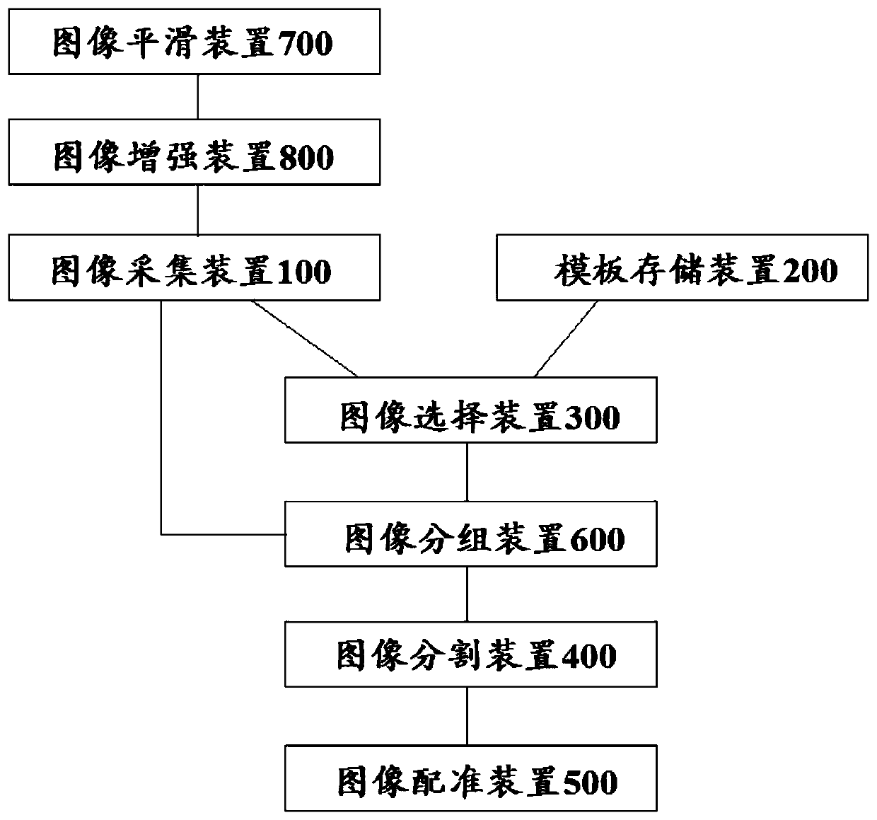
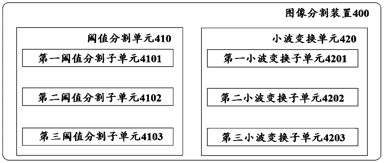
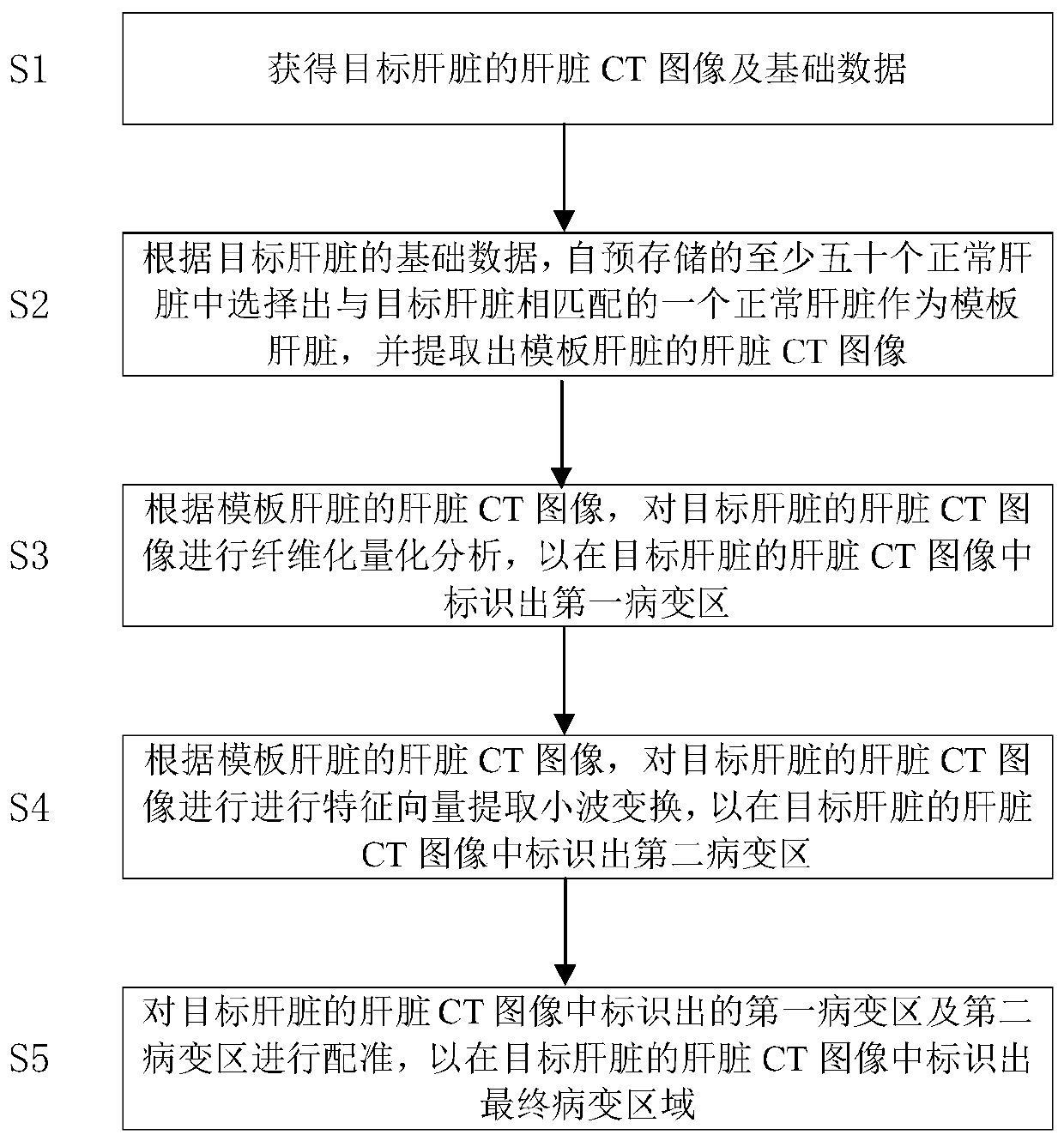
[0026] Please refer to figure 1, as a non-limiting implementation, the system for automatically extracting lesion regions from three-dimensional liver CT images of the present invention includes: an image acquisition device 100 , a template storage device 200 , an image selection device 300 , an image segmentation device 400 and an image registration device 500 .

[0027] The image acquisition device 100 is used to acquire liver CT images of the target liver. The template storage device 200 stores the basic data of at least fift...


PUM


Abstract

The invention discloses a liver three-dimensional CT image lesion area automatic extraction system and method. The system comprises an image acquisition device, a template storage device, an image selection device, an image segmentation device and an image registration device, the image collection device is used for obtaining a liver CT image of a target liver. The template storage device is usedfor storing basic data of at least fifty normal livers and liver CT images. The image selection device is used for selecting a normal liver matched with the target liver as a template liver; the imagesegmentation device is used for carrying out pixel gray difference analysis and texture feature vector extraction wavelet transformation on the liver CT image of the target liver according to the liver CT image of the template liver so as to identify a first lesion area and a second lesion area; and the image registration device is used for registering the first lesion area and the second lesionarea so as to identify a final lesion area in the liver CT image of the target liver.

Description

technical field [0001] The invention relates to a medical image processing method and system, in particular to a liver three-dimensional image processing method and system. Background technique [0002] With the development and maturity of modern medical imaging technology, modern medical equipment can help doctors diagnose patients' internal organ or tissue lesions without surgery. Medical images such as computed tomography (CT) and magnetic resonance imaging (magnetic resonance MR) can generate data representing the information of internal organs of the human body, and play an important role in the diagnosis of diseases. [0003] At present, the diagnosis of liver diseases mainly relies on medical imaging, such as liver CT. At the same time, CT has a high degree of spatial and density resolution, and is especially specific for the display of fat, calcification and water in lesions. Therefore, how to use liver CT images to determine the diseased area of ​​the liver has bec...

Claims


Application Information

Patent Timeline
IPC IPC(8): G06T7/00G06T7/11G06T7/136G06T7/33
CPCG06T7/0012G06T7/11G06T7/136G06T7/33G06T2207/10081G06T2207/30056
Inventor 朱呈瞻董蒨魏宾董冰子房丹夏楠王菲菲王凤娇楚宏硕
Owner THE AFFILIATED HOSPITAL OF QINGDAO UNIV
Why Eureka
Try Eureka