Automatic detection method for various retinal focuses in retinal OCT image

An automatic detection and retinal technology, applied in image enhancement, image analysis, image data processing, etc., can solve the problem of only quantitative analysis of a certain type of lesion, unable to quantitatively analyze the lesion area, unsatisfactory algorithm robustness and accuracy, etc. problems to achieve ideal robustness and accuracy

Pending Publication Date: 2019-08-20
江西比格威医疗科技有限公司
View PDF7 Cites 14 Cited by
  • Summary
  • Abstract
  • Description
  • Claims
  • Application Information

AI Technical Summary

Problems solved by technology

Most of the existing algorithms can only be used for the classification of certain types of lesions, but cannot quantitatively analyze the lesion area; or can only quantitatively analyze a certain type of lesions, and the robustness and accuracy of the algorithm are not ideal

Method used


Image

  • Automatic detection method for various retinal focuses in retinal OCT image
  • Automatic detection method for various retinal focuses in retinal OCT image
  • Automatic detection method for various retinal focuses in retinal OCT image

Examples


Embodiment Construction

[0036] The present invention will be further described below in conjunction with specific embodiments. The following examples are only used to illustrate the technical solution of the present invention more clearly, but not to limit the protection scope of the present invention.

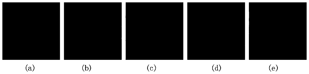
[0037] The retinal lesions detected and segmented by the embodiment of the present invention include 5 types, namely: serous pigment epithelial detachment, subretinal fluid, drusen, choroidal neovascularization and macular hole, such as image 3 shown. in: image 3 (a) An illustration of serous pigment epithelial detachment (PED). The OCT image shows a hemispherical bulge of retinal pigment epithelium light bands, which are mostly early manifestations or accompanying symptoms of age-related macular degeneration. image 3 (b) An illustration of a subretinal fluid (SRF). In an OCT image, the interior of the subretinal fluid presents a black dark reflection signal. Its components come from retinal deg...


PUM


Abstract

The invention provides an automatic detection method for various retinal focuses in a retinal OCT image. The automatic detection method comprises the following steps: acquiring a retinal 3D OCT image;carrying out detection and preliminary segmentation on the retina 3D OCT image by adopting multiple trained retina focus detection and segmentation models to obtain a focus area subjected to preliminary segmentation; and correcting the preliminarily segmented lesion area to obtain an accurate retinal lesion area. According to the automatic detection method, the retinal focus area in the retinal OCT image can be accurately detected, and various retinal focuses in the retinal OCT image can be quantitatively analyzed, and ideal robustness and accuracy are achieved.

Description

technical field [0001] The invention relates to an automatic detection method for multiple retinal lesions in retinal OCT images, and belongs to the technical field of image processing and analysis. Background technique [0002] Serous pigment epithelial detachment, subretinal fluid, choroidal neovascularization, and drusen are important clinical manifestations of severe retinal diseases such as age-related macular degeneration and diabetic macular edema. Macular holes are also serious retinal diseases. These disorders can cause vision loss, physical blind spots, and even blindness. Therefore, accurate detection and segmentation of lesion areas is of great significance for obtaining precise treatment and reducing treatment costs. Currently available detection methods for retinal diseases / lesions on retinal OCT images have limitations. Most of the existing algorithms can only be used for the classification of certain types of lesions, but cannot quantitatively analyze the l...

Claims


Application Information

Patent Timeline
Patent Type & Authority Applications(China)
IPC IPC(8): G06T7/00G06T7/10A61B3/10
CPCG06T7/0012G06T7/10A61B3/102G06T2207/10101G06T2207/20081G06T2207/20084G06T2207/30041
Inventor 陈新建管丽玲俞凯
Owner 江西比格威医疗科技有限公司
Why Eureka
Try Eureka