
Medical image processing method, system and device and computer readable medium

A medical image and processing method technology, applied in the field of medical image processing methods and systems, can solve problems such as difficult to locate lesions, low PET resolution, and inability to provide structural information of patients

Active Publication Date: 2019-08-23
SHANGHAI UNITED IMAGING HEALTHCARE
View PDF4 Cites 11 Cited by
  • Summary
  • Abstract
  • Description
  • Claims
  • Application Information

AI Technical Summary

Problems solved by technology

However, the resolution of PET is low and cannot provide structural information of the patient, making it difficult to locate the lesion only relying on PET images

Method used


Image

  • Medical image processing method, system and device and computer readable medium
  • Medical image processing method, system and device and computer readable medium
  • Medical image processing method, system and device and computer readable medium

Examples


Embodiment Construction

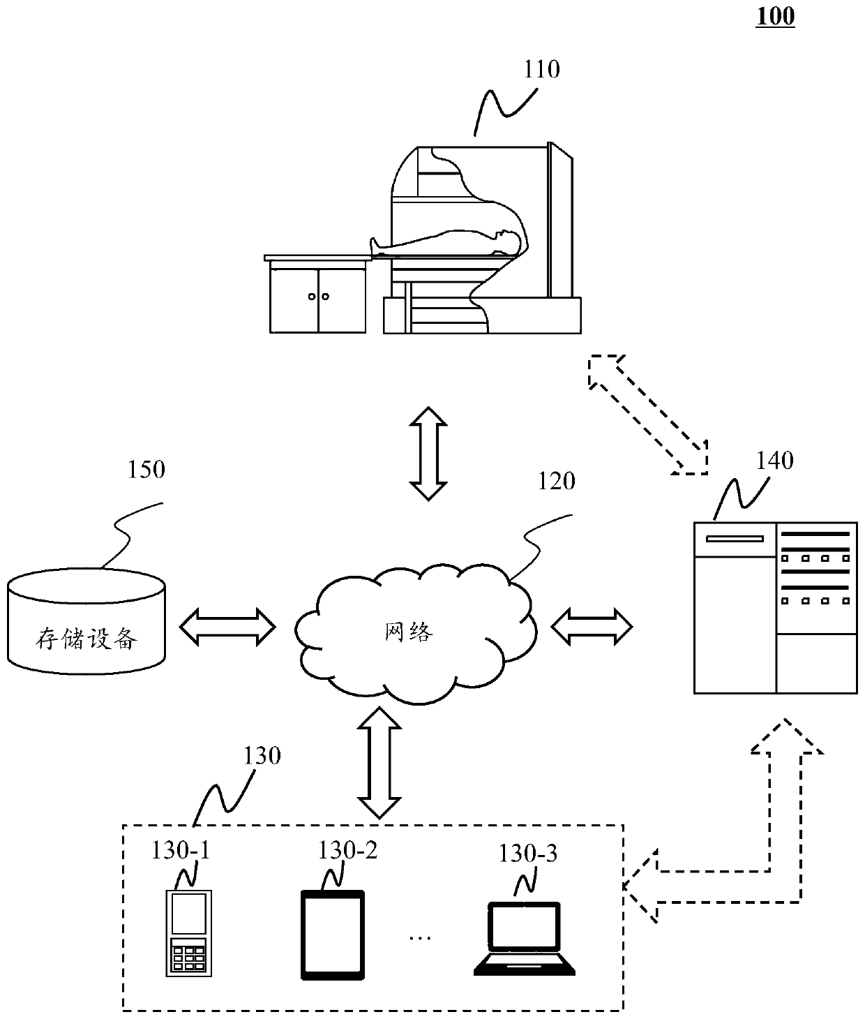
[0026] In order to more clearly illustrate the technical solutions of the embodiments of the present application, the following briefly introduces the drawings that need to be used in the description of the embodiments. Obviously, the accompanying drawings in the following description are only some examples or embodiments of the present application, and those skilled in the art can also apply the present application to other similar scenarios. Unless otherwise apparent from context or otherwise indicated, like reference numerals in the figures represent like structures or operations.

[0027] It should be understood that "system", "device", "unit" and / or "module" as used herein is a method used to distinguish different components, elements, parts, parts or assemblies of different levels. However, the words may be replaced by other expressions if other words can achieve the same purpose.

[0028] As indicated in this application and claims, the terms "a", "an", "an" and / or "t...


PUM


Abstract

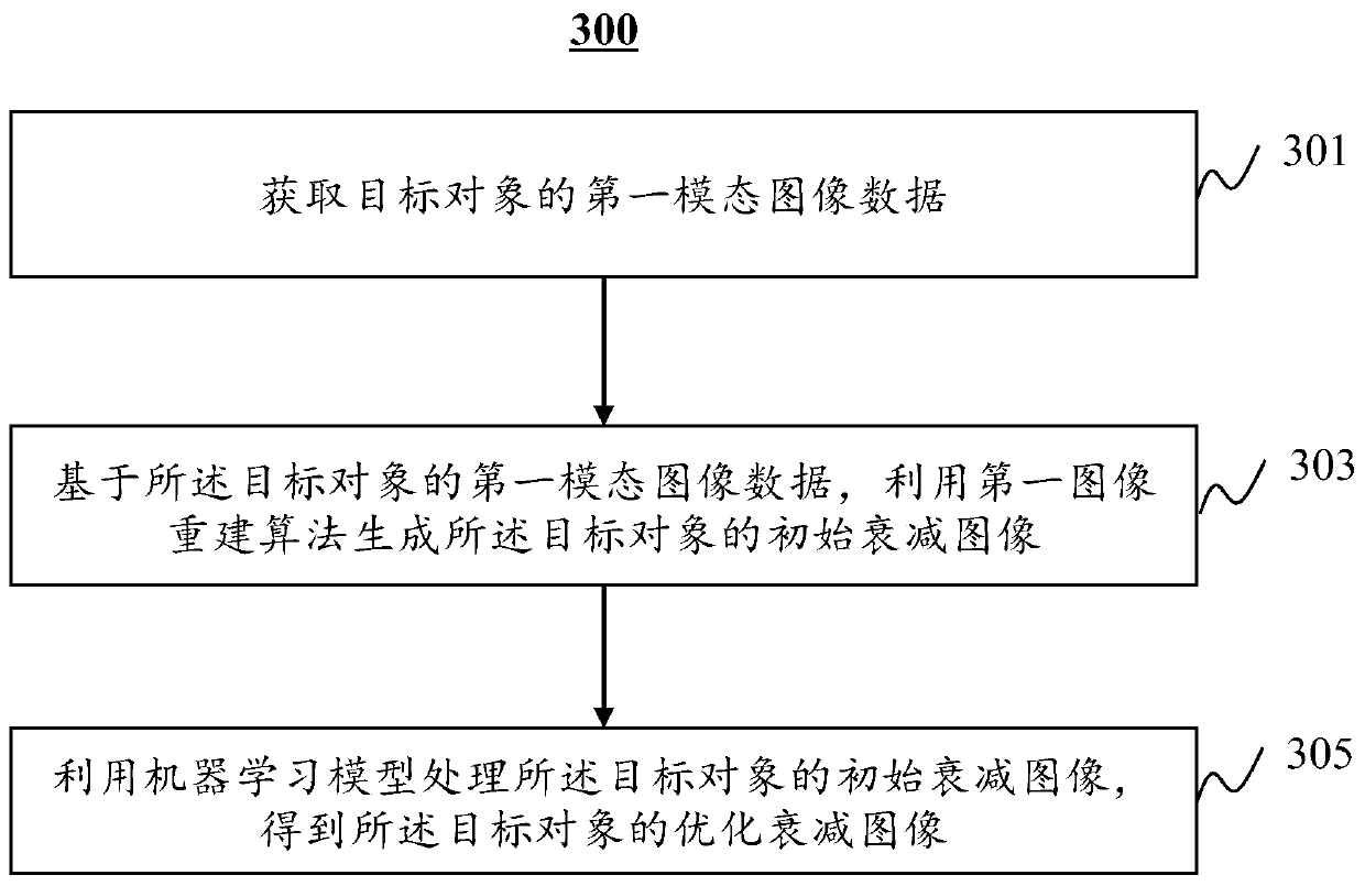
The invention provides a medical image processing method, system and device and a computer readable storage medium. The method comprises the steps that first modal image data of a target object is acquired; based on the first modal image data of the target object, a first image reconstruction algorithm is used for generating an initial attenuation image of the target object; a machine learning model is used for processing the initial attenuation image of the target object to obtain an optimized attenuation image of the target object; according to the optimized attenuation image of the target object, attenuation correction is carried out on the first modal image data of the target object to obtain a first modal image after attenuation correction, or according to the optimized attenuation image of the target object, a second modal image of the target object is determined. The medical image processing method, system and device and the computer readable medium can be used for obtaining themore accurate attenuation image under the situation that a patient is free of additional scanning.

Description

technical field [0001] The present application relates to the field of image processing, in particular to a medical image processing method and system. Background technique [0002] Positron Emission Tomography (PET) is the highest level of nuclear medicine imaging technology today, and it has shown excellent performance in the fields of oncology, cardiovascular disease, nervous system disease research, and new drug development research. PET reconstructs the gamma photon pairs of tracers in the human body to produce images reflecting the metabolic function of the scanned object. However, the resolution of PET is low and cannot provide structural information of the patient, making it difficult to locate the lesion only relying on PET images. Therefore, it is necessary to provide a method for obtaining a structural image of a target object from positron emission tomography data. Contents of the invention [0003] One aspect of the present application provides a medical ima...

Claims


Application Information

Patent Timeline
Patent Type & Authority Applications(China)
IPC IPC(8): A61B6/03
CPCA61B6/037A61B6/5211
Inventor 冯涛龚南杰
Owner SHANGHAI UNITED IMAGING HEALTHCARE
Learn More