3D medical image recognition and segmentation method based on Unet and LSTM

A medical imaging and imaging technology, applied in the field of medical image recognition and segmentation, can solve the problems of lack of contextual connection, low computational efficiency of 3D neural network, large amount of 3D medical imaging data, etc., and achieve good contextual relationship mining and segmentation result quality. high effect

Inactive Publication Date: 2019-11-05
EAST CHINA NORMAL UNIV
View PDF3 Cites 9 Cited by
  • Summary
  • Abstract
  • Description
  • Claims
  • Application Information

AI Technical Summary

Problems solved by technology

3D medical images have a large amount of data, and the processing and calculation efficiency of 3D neural networks is low, while the segmentation results of 2D images lack good contextual connections in the z-axis dimension

Method used


Image

  • 3D medical image recognition and segmentation method based on Unet and LSTM
  • 3D medical image recognition and segmentation method based on Unet and LSTM
  • 3D medical image recognition and segmentation method based on Unet and LSTM

Examples


Embodiment

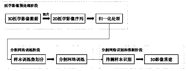
[0032] Taking brain tumor MRI images as an example, the steps are as follows:

[0033] Medical image preprocessing stage:

[0034] First, read 3D format medical images from MRI instruments that support 3D format scanning. The original image is recorded as x, x is a three-dimensional matrix, and the size is length m, width n, height k, and the dimension with rich information is selected as the z axis (such as: top view ), for example: slice the 3D image along high k to form a 2D image sequence, namely:

[0035] x=[x 1 ,x 2 ,...,x i ,...,x k ]

[0036] x 1 ,x 2 ,...,x i ,...,x k A sequence of slices for each 2D image, x i is the i-th 2D image slice, the length is m and the width is n, and the sequence length is k. Similarly, the segmentation annotation y of the 3D image is also processed to form a sequence of 2D segmentation annotation slices with the same shape as x:

[0037] y=[y 1 ,y 2 ,...,y i ,...,y k ]

[0038] the y i Annotate slices for the i-th 2D segm...


PUM


Abstract

The invention discloses a 3D medical image recognition and segmentation method based on Unet and LSTM. The method comprises the steps: a medical image preprocessing stage: reading a medical image in a3D format, enabling the medical image to be decomposed into a 2D image sequence in a z-axis direction, and carrying out the z-score normalization processing of image data in a 2D level; a segmentation network training stage: using the normalized 2D image sequence samples to divide a training set for training, independently taking out the output of the intermediate layer of the U-net as an intermediate variable sequence, and using the intermediate variable sequence to train an LSTM network; and a segmentation network identification and inference stage: inputting the sample into the network toobtain pixel-level segmentation output, and combining output sequences into a 3D matrix to obtain a final result. In the aspect of 3D medical image recognition, the mode that the 3D medical image is segmented into the 2D sequence and the intermediate variable is processed in combination with the cyclic network is adopted. The calculated amount is reduced, and the efficiency of recognition work isimproved.

Description

technical field [0001] The present invention relates to the identification and segmentation technology of medical images based on neural network, in particular to a method for identification and segmentation of 3D medical images based on U-shaped network (Unet) and long-short-term memory network (Long Short-Term Memory, LSTM). Background technique [0002] In daily medical work and clinical trials, the application of medical imaging is very popular, and the method of artificial neural network has also provided great help in medical imaging. Generally, the significance of using medical imaging lies in measurement and recording, so its data contains strong positioning characteristics (such as tumors, images of fetuses, etc.), and related precision instruments use various imaging principles such as optics and sound waves. Visualize the organs and tissues in the human body, so as to make judgments on the physical properties, structural characteristics and current working status ...

Claims


Application Information

Patent Timeline
Patent Type & Authority Applications(China)
IPC IPC(8): G06K9/00G06K9/34G06K9/62G06N3/04G06T7/11G16H30/40
CPCG06T7/11G16H30/40G06V20/647G06V20/64G06V10/267G06N3/045G06F18/214
Inventor 朱思涵浦剑
Owner EAST CHINA NORMAL UNIV
Why Eureka
Try Eureka