Isolated culture method for tumor-specific T cells and product obtained through method

A tumor-specific, separation and culture technology, applied in the field of separation and culture methods and products obtained therefrom, achieves the effects of convenient material collection, convenient blood collection, and short time required.

Inactive Publication Date: 2019-11-15
HENAN CANCER HOSPITAL
View PDF9 Cites 8 Cited by
  • Summary
  • Abstract
  • Description
  • Claims
  • Application Information

AI Technical Summary

Problems solved by technology

But this method can only get CD4 + T lymphocytes

Method used


Image

  • Isolated culture method for tumor-specific T cells and product obtained through method
  • Isolated culture method for tumor-specific T cells and product obtained through method
  • Isolated culture method for tumor-specific T cells and product obtained through method

Examples


Embodiment 1

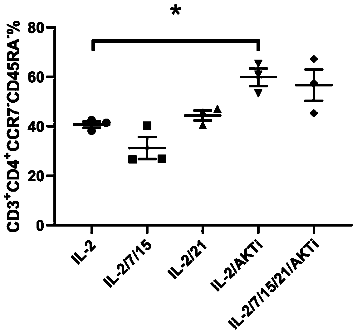
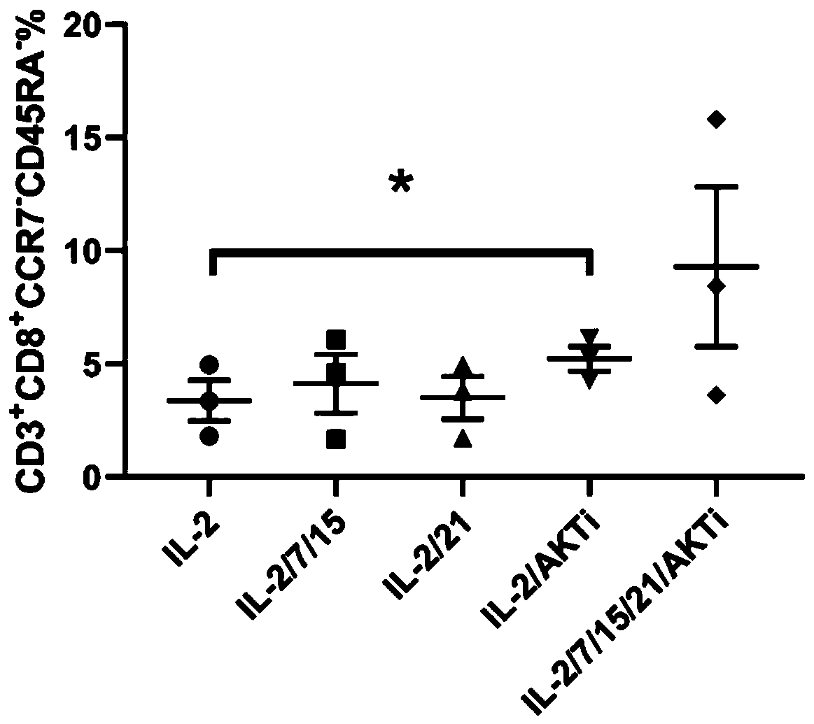
[0055] Influence of Factor Types in Culture Medium on the Proportion of Effector Memory T Lymphocytes in Cell Final Products

[0056] This example explores the effects of five different culture methods on effector memory T cells (specific phenotype: CD3) in the final cell product. + CD8 + CD45RA - CCR7 - and CD3 + CD4 + CD45RA - CCR7 - ) ratio, its specific operation includes the following steps:

[0057] (1) Take the peripheral blood of kidney cancer patients who have been treated with PD-1 antibody within 3 weeks;

[0058] (2) The peripheral blood obtained in step (1) is separated by the Ficoll density gradient method to obtain mononuclear cells;

[0059] (3) Incubate the mononuclear cells obtained in step (2) with biotinylated anti-human IgG4 antibody at 25°C for 20 minutes, then add magnetic beads coated with anti-biotin antibody and incubate at 25°C for 10 minutes, Magnetic separation of mononuclear cells positive for PD-1;

[0060] (4) The PD-1 positive mononuc...

Embodiment 2

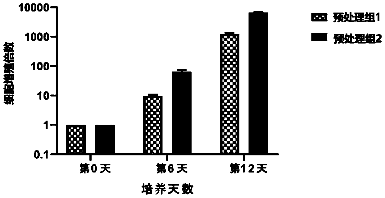
[0063] Effects of two different PD-1 antibody pretreatment methods on the proliferation ability of PD-1 positive cells

[0064] This example explores the effects of two different PD-1 antibody pretreatment methods on the expansion speed of isolated tumor-specific T cells. In this example, two experiments were carried out on samples, one of which was peripheral blood from patients with cervical cancer. (sample 1), and the second is the peripheral blood (sample 2) of a patient with malignant melanoma. The specific operation includes the following steps:

[0065] (1) Collect peripheral blood from patients with cervical cancer and malignant melanoma before and after PD-1 antibody drug treatment (within 3 weeks). Add PD-1 antibody to the peripheral blood of patients who have not been treated with PD-1 antibody drug to make the final concentration 0.5 μg / mL, and incubate at 4°C for 0.5h, as pretreatment group 1; The patient's peripheral blood after drug treatment is directly used i...

Embodiment 3

[0070] Evaluation test for tumor specificity of isolated T cells (sample is peripheral blood of renal cancer patients)

[0071] This example evaluates the ability of isolated T cells to recognize autologous tumor cells, using IFN-γ-secreting cells to account for CD8 + and CD4 + The proportion of cells (ie, CD8 + IFN-γ + %, CD4 + IFN-γ + %) for characterization, the higher the ratio, the higher the tumor specificity of T cells.

[0072] The specific operation includes the following steps:

[0073] (1) The test is divided into three groups, which are unsorted group, PD-1 negative group and PD-1 positive group;

[0074] (2) Take peripheral blood from kidney cancer patients and add PD-1 antibody for incubation (pretreatment);

[0075] (3) The peripheral blood obtained in step (2) is separated by the Ficoll density gradient method to obtain mononuclear cells;

[0076](4) Incubate the mononuclear cells obtained in step (3) with biotinylated anti-human IgG4 antibody at 25°C f...


PUM


Abstract

The invention relates to an isolated culture method for tumor-specific T cells and a product obtained through the method. The isolated culture method comprises the steps that firstly, peripheral bloodof a tumor patient is pretreated, mononuclear cells are separated from the peripheral blood, the mononuclear cells with positive PD-1 are separated out, finally, the mononuclear cells with positive PD-1 are amplified, and the tumor-specific T cells are obtained. An initial material involved by the isolated culture method for the tumor-specific T cells is the peripheral blood of the tumor patient,tumor tissue or malignant pleural effusion and ascites of the patient do not need to be used as the material, the material is more conveniently collected, blood sampling can be conveniently conductedon almost all tumor patients, and no large wounds are caused; the finally-obtained T cells are high in tumor specificity and comprise T cells with positive CD4 and T cells with positive CD8; the method is simple in operation process and short in required time.

Description

technical field [0001] The invention belongs to the technical field of cell culture, and in particular relates to a method for separating and culturing tumor-specific T cells and products obtained therefrom. Background technique [0002] Malignant tumors seriously endanger human life and health, and its mortality rate ranks first among various diseases. The three existing conventional treatment methods for tumors (i.e. surgery, radiotherapy and chemotherapy) have made significant contributions to improving the survival rate of cancer patients in the past few decades, but they have not been able to solve the problem of tumor metastasis and recurrence. However, the survival rate of cancer patients has not improved significantly in recent years. The three conventional treatments have encountered curative effect bottlenecks, and there is an urgent need to find new methods that can break through the bottleneck of tumor therapeutic efficacy. [0003] Tumor-specific T lymphocytes ...

Claims


Application Information

Patent Timeline
Patent Type & Authority Applications(China)
IPC IPC(8): C12N5/0783
CPCC12N5/0636C12N2509/00C12N2501/2302C12N2501/2307C12N2501/2315C12N2501/2321C12N2501/727C12N2533/52C12N2501/515
Inventor 李铁鹏高全立王瑶
Owner HENAN CANCER HOSPITAL
Why Eureka
Try Eureka