Isolated culture method of tumor-specific T cells and product obtained by isolated culture method
A tumor-specific, separation and culture technology, applied in the field of separation and culture methods and products obtained therefrom, to achieve the effects of convenient blood collection, short time required and high tumor specificity
- Summary
- Abstract
- Description
- Claims
- Application Information
AI Technical Summary
Problems solved by technology
Method used
Image
Examples
Embodiment 1
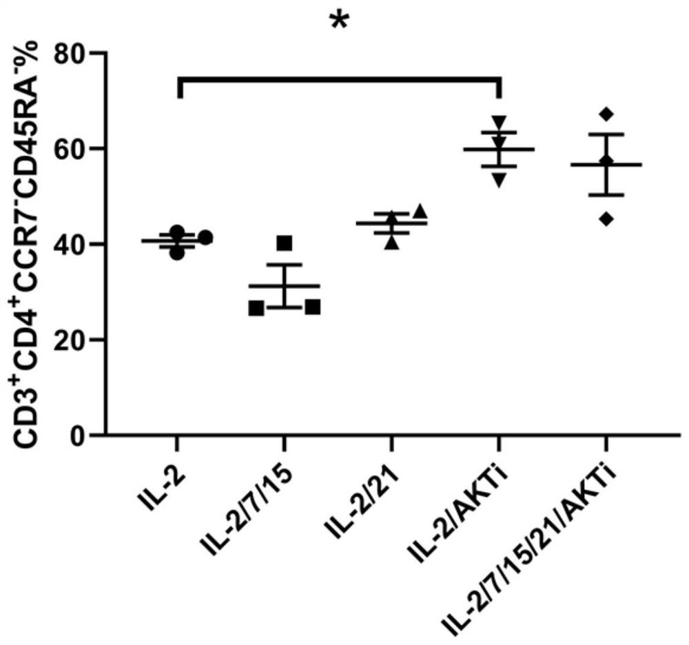
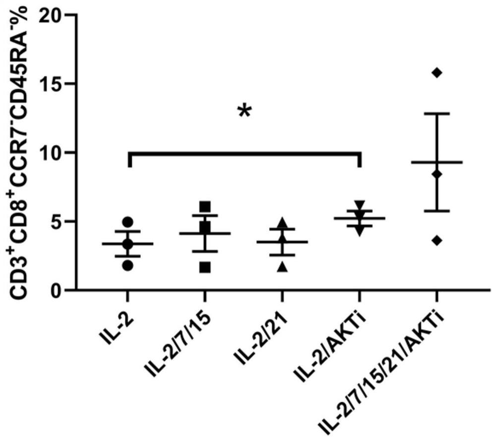
[0056] Effect of Factor Types in the Culture Medium in Example 1 on the Effector Memory T Lymphocyte Ratio in Cell Final Products
[0057] This example explores the effects of five different culture methods on effector memory T cells (specific phenotype: CD3) in the final cell product. + CD8 + CD45RA - CCR7 - and CD3 + CD4 + CD45RA - CCR7 - ) ratio, its specific operation includes the following steps:
[0058] (1) Take the peripheral blood of kidney cancer patients who have been treated with PD-1 antibody within 3 weeks;
[0059] (2) The peripheral blood obtained in step (1) is separated by the Ficoll density gradient method to obtain mononuclear cells;
[0060] (3) The mononuclear cells obtained by step (2) are separated and used at a concentration of 10 7 Mononuclear cells were co-incubated with 2.5 μg of biotinylated anti-human IgG4 antibody for 20 minutes at 25°C, then added anti-biotin antibody-coated magnetic beads and incubated for 10 minutes at 25°C. Magnetic ...
Embodiment 2
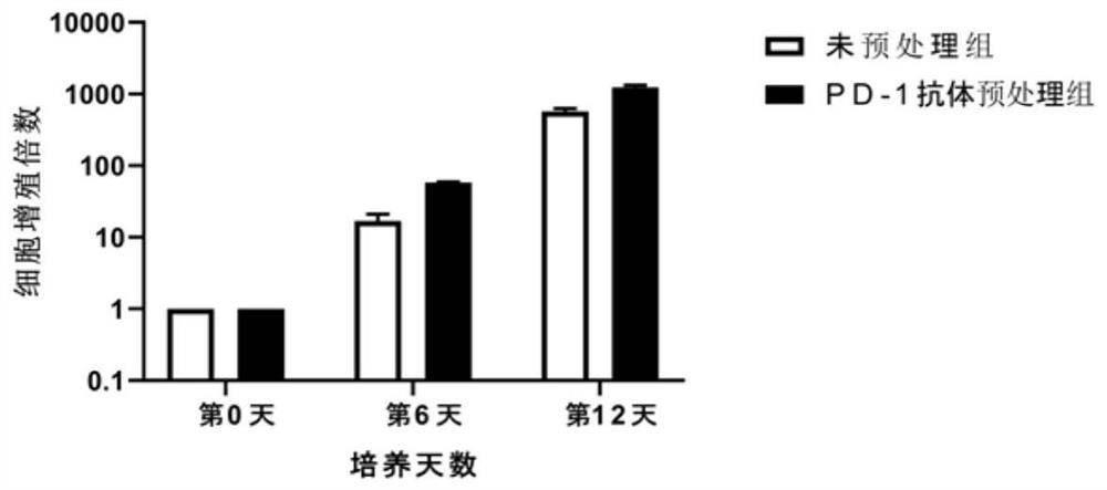
[0063] Example 2 Effects of Two Different PD-1 Antibody Pretreatment Methods on the Proliferative Ability of PD-1 Positive Cells
[0064] This example explores the effects of two different PD-1 antibody pretreatment methods on the expansion speed of isolated tumor-specific T cells. In this example, two experiments were carried out on samples, one of which was peripheral blood from patients with cervical cancer. (sample 1), and the second is the peripheral blood (sample 2) of a patient with malignant melanoma. The specific operation includes the following steps:
[0065] (1) Collect peripheral blood from patients with cervical cancer and malignant melanoma before and after PD-1 antibody drug treatment (within 3 weeks). Add PD-1 antibody to the peripheral blood of patients who have not been treated with PD-1 antibody drug to make the final concentration 0.5 μg / mL, and incubate at 4°C for 0.5h, as pretreatment group 1; The patient's peripheral blood after drug treatment is direc...
Embodiment 3
[0069] Example 3 Evaluation Test of the Tumor Specificity of the Isolated T Cells (The Sample Is the Peripheral Blood of Renal Cancer Patients)
[0070] This example evaluates the ability of isolated T cells to recognize autologous tumor cells, using IFN-γ-secreting cells to account for CD8 + and CD4 + The proportion of cells (ie, CD8 + IFN-γ + %, CD4 + IFN-γ + %) for characterization, the higher the ratio, the higher the tumor specificity of T cells.
[0071] The specific operation includes the following steps:
[0072] (1) The test is divided into three groups, which are unsorted group, PD-1 negative group and PD-1 positive group;
[0073] (2) Take peripheral blood from kidney cancer patients, add PD-1 antibody to make the final concentration 0.5 μg / mL and incubate (pretreatment);
[0074] (3) The peripheral blood obtained in step (2) is separated by the Ficoll density gradient method to obtain mononuclear cells;
[0075] (4) The mononuclear cells obtained by step (3) ...
PUM
| Property | Measurement | Unit |
|---|---|---|
| concentration | aaaaa | aaaaa |
Abstract
Description
Claims
Application Information
 - R&D
- Intellectual Property
- Life Sciences
- Materials
- Tech Scout
- Unparalleled Data Quality
- Higher Quality Content
- 60% Fewer Hallucinations
Browse by: Latest US Patents, China's latest patents, Technical Efficacy Thesaurus, Application Domain, Technology Topic, Popular Technical Reports.
© 2025 PatSnap. All rights reserved.Legal|Privacy policy|Modern Slavery Act Transparency Statement|Sitemap|About US| Contact US: help@patsnap.com


