
A method for preparing in situ tissue engineering blood vessels by a composite process

A technology of tissue engineering and composite technology, applied in the field of biomedical tissue engineering, to achieve the effect of simple and feasible removal steps, guaranteed mechanical properties, and increased heparin molecular binding sites

Active Publication Date: 2022-01-07
SHANGHAI UNIV
View PDF6 Cites 0 Cited by
  • Summary
  • Abstract
  • Description
  • Claims
  • Application Information

AI Technical Summary

Problems solved by technology

Therefore, the material selection and preparation process of tissue engineering blood vessels still need to be developed and improved.

Method used


Image

  • A method for preparing in situ tissue engineering blood vessels by a composite process
  • A method for preparing in situ tissue engineering blood vessels by a composite process
  • A method for preparing in situ tissue engineering blood vessels by a composite process

Examples


Embodiment 1

[0036] like Figure 5 As shown, a method for preparing in situ tissue engineered blood vessels by a composite process comprises the following steps:

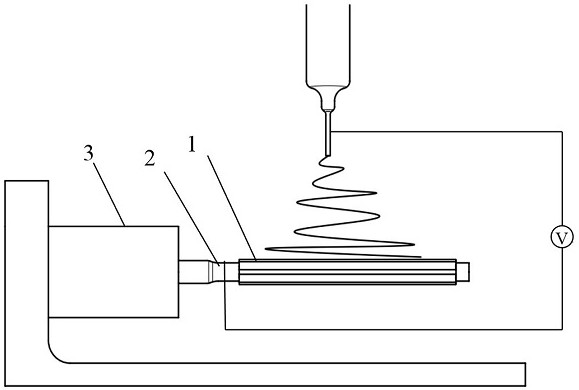
[0037] a. 3D printing water-soluble materials to prepare stent collection rods: the structure is a cylindrical hollow structure, such as figure 1 As shown: the inner diameter is 3.5mm, the outer diameter is 4.5mm, and the outer surface of the cylinder is designed with 8 ridges. The size of the raised structure is 50mm in length, 0.4mm in width, and 0.2mm in height. The collection rod is made of polyvinyl alcohol, a water-soluble polymer material Shaped by FDM printing. The nozzle temperature of the printer is 160°C, the bed temperature is 60°C, the nozzle moving speed is 20mm / s, and the layer thickness is set to 0.15mm.

[0038] b. The inner layer of tissue engineering blood vessels was prepared by electrospinning process, such as figure 2 Shown: To increase the number of end groups available for biomolecular attachment, pol...

Embodiment 2

[0045] This embodiment is basically the same as Embodiment 1, and the special features are:

[0046] In this embodiment, a method for preparing in situ tissue engineered blood vessels by a composite process is to prepare ridges of different widths and numbers on the outside of the stent collection rod to cause patterning of the inner layer of the stent, including the following steps:

[0047] a. 3D printing water-soluble materials to prepare vascular stent collection rods: design vascular stent collection rods: its structure is a cylindrical hollow structure with an inner diameter of 4mm and an outer diameter of 5mm, and 6 ridges are designed on the outer surface of the cylinder, and the size of the raised structure is long 50mm, 0.5mm wide, and 0.3mm high, the collection rod is made of water-soluble polymer material polyvinyl alcohol and formed by FDM printing. The nozzle temperature of the printer is 160°C, the bed temperature is 60°C, the nozzle moving speed is 20mm / s, and ...


PUM


Abstract

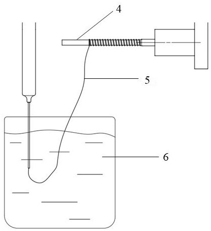
The invention relates to a method for preparing in situ tissue engineering blood vessels by a composite process. Combining rapid prototyping, electrospinning and wet spinning processes to prepare a three-layer composite vascular scaffold for in situ tissue engineering, which is similar to natural blood vessel structure; its inner layer is prepared by 3D printing and electrospinning technology with axial A tubular scaffold with grooves; the middle layer is a tubular scaffold with circumferentially arranged fibers prepared by wet spinning; the outer layer is a tubular scaffold composed of disordered fibers that can cover the inner layer and the middle layer prepared by electrospinning. The vascular stent prepared by the present invention is also loaded with vascular endothelial cell growth factor through heparinization, which can recruit cells in the host body, reduce the chance of infection in vitro culture, and realize the preparation of in situ tissue engineering blood vessels, which has broad application in the field of tissue engineering blood vessels Prospects.

Description

technical field [0001] The invention relates to the technical field of biomedical tissue engineering, in particular to a method for preparing blood vessels for tissue engineering in situ by a composite process. Background technique [0002] With the acceleration of population aging, many diseases related to human blood vessels, such as atherosclerosis and coronary heart disease, are on the rise. When the human body's own blood vessels cannot normally supply blood to the human body due to factors such as disease and trauma, surgical operations such as repair, intravascular stents, or autologous vein grafting should be used to treat the damaged blood vessels. Generally, the saphenous vein is considered to be a good substitute for autologous blood vessels in small-diameter vascular reconstruction, but some patients cannot use autologous veins due to their own diseases or other reasons, so using tissue engineered blood vessels as a substitute for tissue vascular repair has becom...

Claims


Application Information

Patent Timeline
Patent Type & Authority Patents(China)
IPC IPC(8): A61L27/40A61L27/18A61L27/34A61L27/50A61L27/54
CPCA61L27/18A61L27/34A61L27/507A61L27/54A61L27/50C08L67/04
Inventor 陈箫姚远刘随红张亦弛
Owner SHANGHAI UNIV
Learn More