
A Method of 3D Reconstruction of Medical Images Based on Neighborhood Topology
A medical image and three-dimensional reconstruction technology, applied in the field of combining medicine and engineering science, can solve problems such as poor repair effect, reduce the amount of calculation, improve the speed of online three-dimensional reconstruction, and improve the adaptability.
Patent Information
- Authority / Receiving Office
- CN · China
- Patent Type
- Patents(China)
- Current Assignee / Owner
- Publication Date
- 2021-05-25
Smart Images
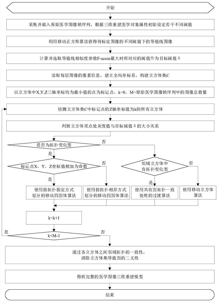
Figure 1 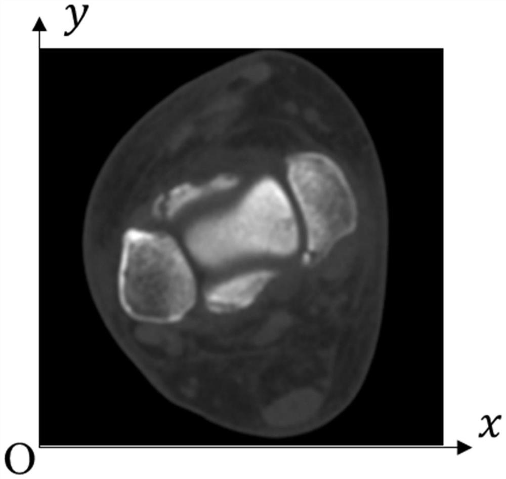
Figure 2 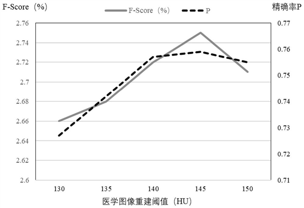
Figure 3
Abstract
Description
technical field
[0001] The invention belongs to the field of combination of medicine and engineering science, and relates to a processing method of a geometric structure model in the process of medical image processing and three-dimensional reconstruction. Background technique
[0002] A medical image sequence usually refers to a sliced parallel image sequence describing a medical object. The acquisition method can be computerized tomography (Computed Tomography, CT), ultrasonic imaging (Ultrasonic Imaging, UI), magnetic resonance imaging (Magnetic Resonance Imaging, MRI), Nuclear Medicine Imaging (NMI), Doppler Flow Imaging (DFI), Positron Emission Tomography (PET), Single Photon Emission Computed Tomography (Single PhotonEmission Computed Tomography, SPECT) and so on. The precision and accuracy of reconstruction of 3D medical tissue from 2D medical image sequences directly determine the effect of medical analysis and diagnosis. The reconstructed three-dimensional geome...
Examples
specific Embodiment
[0064] Step 1: Taking the human radial styloid process and ulnar styloid process as three-dimensional reconstruction medical objects, initially set the threshold as the gray value of the periosteum and several thresholds similar to the gray value, and select 130HU, 135HU, 140HU, 145HU and 150HU is used as the initial threshold;
[0065] Step 2: Collect and load the original skeletal medical image frame sequence of the 3D reconstructed medical object, select the 121-layer CT image, and select the CT image to be calibrated in the original skeletal medical image frame sequence, the CT image contains the complete 3D reconstructed medical object Contour, according to the threshold set in step 1, use the moving square algorithm (Marching Squares, MS) to process the CT image to obtain the contour and its contour image, and then compare the contours under different thresholds, as follows:
[0066] Step 2.1: use the Canny edge detection operator to extract the edges of the radius and u...