Retinal fundus image segmentation method and device

A fundus image and retina technology, which is applied in image analysis, image enhancement, medical image and other directions, can solve the problem of poor segmentation accuracy of blood vessels and optic disc in retinal fundus images, and achieve the effect of solving the problem of insufficient labeling samples and accurate segmentation.

Inactive Publication Date: 2020-01-17
UNIV OF SCI & TECH BEIJING
View PDF3 Cites 16 Cited by
  • Summary
  • Abstract
  • Description
  • Claims
  • Application Information

AI Technical Summary

Problems solved by technology

[0003] The technical problem to be solved by the present invention is to provide a retinal fundus image segmentation method and device to solve the problem that the deep learning method based on a small number of labeled samples in the prior art will lead to poor segmentation accuracy of retinal fundus image vessels and optic discs question

Method used


Image

  • Retinal fundus image segmentation method and device
  • Retinal fundus image segmentation method and device
  • Retinal fundus image segmentation method and device

Examples


Embodiment 1

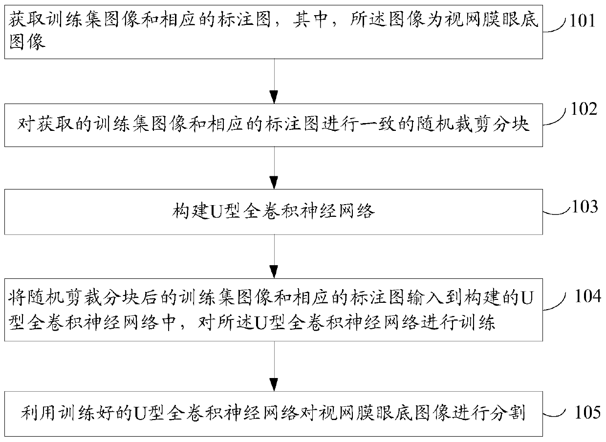
[0042] Such as figure 1 As shown, the retinal fundus image segmentation method provided by the embodiment of the present invention includes:

[0043] S101. Obtain training set images and corresponding labeled images, wherein the images are retinal fundus images;

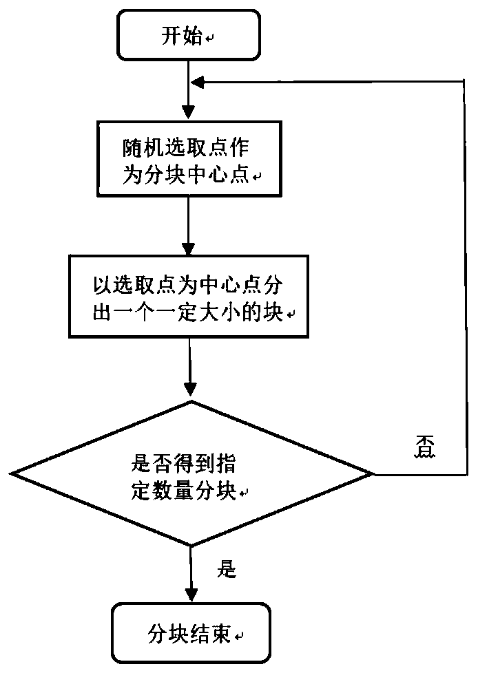
[0044] S102, perform consistent random cropping and segmentation of the acquired training set images and corresponding labeled images;

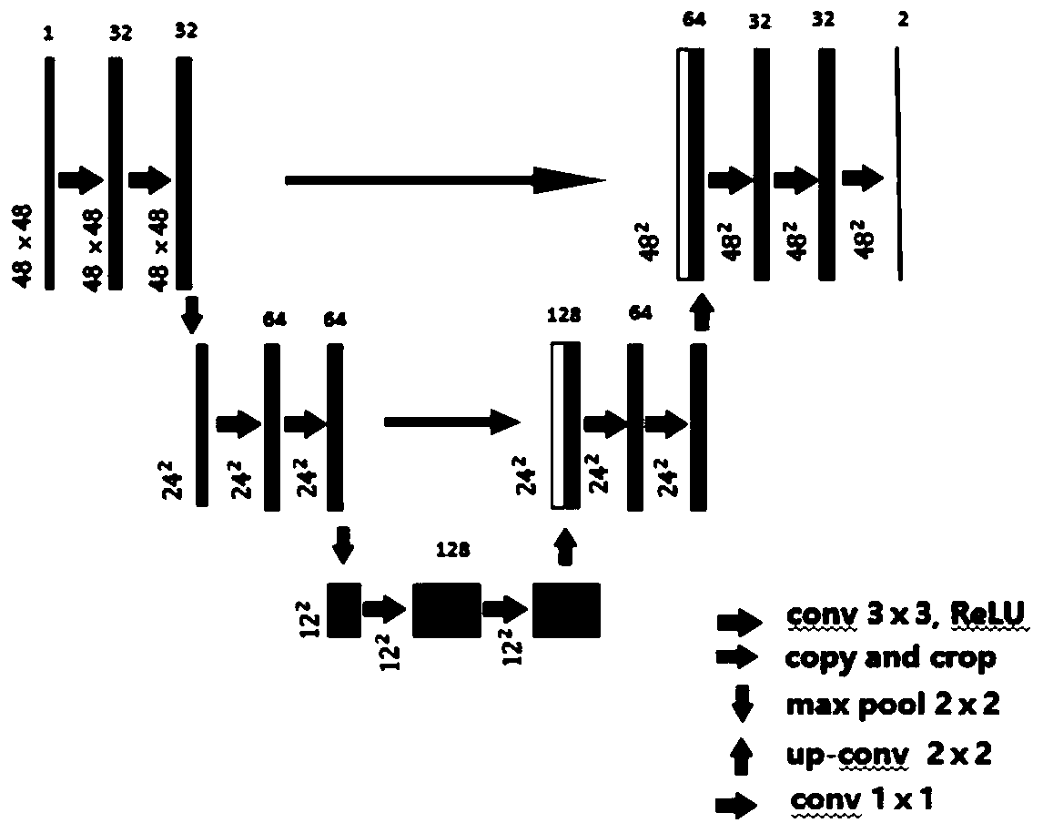
[0045] S103, constructing a U-shaped fully convolutional neural network;

[0046] S104, inputting the training set image after random clipping and segmentation and the corresponding labeled image into the constructed U-shaped fully convolutional neural network, and training the U-shaped fully convolutional neural network;

[0047] S105, using the trained U-shaped fully convolutional neural network to segment the retinal fundus image.

[0048] The retinal fundus image segmentation method described in the embodiment of the present invention obtains a training set image and a corres...


PUM


Abstract

The invention provides a retinal fundus image segmentation method and device, which can realize automatic, efficient and accurate segmentation of retinal fundus image blood vessels and optic disks under the condition of only a small number of labeled samples. The method comprises the steps of acquiring a training set image and a corresponding annotation image, wherein the image is a retinal fundusimage; carrying out consistent random cutting and blocking on the obtained training set image and the corresponding annotation image; constructing a U-shaped full convolutional neural network; inputting the randomly clipped and blocked training set images and the corresponding annotation images into a constructed U-shaped full convolutional neural network, and training the U-shaped full convolutional neural network; and segmenting the retinal fundus image by using the trained U-shaped full convolutional neural network. The invention relates to the field of artificial intelligence and diabeticretina diagnosis.

Description

technical field [0001] The invention relates to the fields of artificial intelligence and diabetic retinal diagnosis, in particular to a retinal fundus image segmentation method and device. Background technique [0002] Since retinal fundus blood vessels are the only blood vessels that can be directly observed in a non-invasive way, information such as blood vessels obtained from retinal fundus image segmentation is an important indicator for the diagnosis of diabetic retinopathy. However, manual segmentation of retinal fundus images is time-consuming and laborious. If the method of deep learning is used, a large amount of labeled data is required to complete it. If the number of labeled samples is small, the accuracy of the segmentation of blood vessels and optic discs in retinal fundus images will be poor. However, the width of fundus blood vessels ranges from 1 pixel to 20 pixels, and there are crossings, branches, and centerline reflections of blood vessels. Manual label...

Claims


Application Information

Patent Timeline
Patent Type & Authority Applications(China)
IPC IPC(8): G06T7/10G06T7/00G06N3/04G06N3/08G16H30/20
CPCG06T7/10G06T7/0014G06N3/08G16H30/20G06T2207/20021G06T2207/20081G06T2207/30041G06N3/045
Inventor 阿孜古丽.吾拉木未忠杰张德政蒋彦钊孙振起李军胡良缘
Owner UNIV OF SCI & TECH BEIJING
Why Eureka
Try Eureka