Method for automatically segmenting mammary gland calcification points based on deep learning

An automatic segmentation and breast calcification technology, which is applied in the field of breast X-ray auxiliary diagnosis, can solve problems such as misdiagnosis or missed diagnosis, difficulty in finding calcification points, etc., and achieve the effect of improving accuracy

Pending Publication Date: 2020-01-31
ZHEJIANG DE IMAGE SOLUTIONS CO LTD
View PDF1 Cites 2 Cited by
  • Summary
  • Abstract
  • Description
  • Claims
  • Application Information

AI Technical Summary

Problems solved by technology

However, the calcification points in the breast are small and distributed in large numbers on the breast tissue. It is difficult to find all the calcification points and some tiny calcification points with a diameter of less than 5mm by manual reading, which can easily lead to misdiagnosis or missed diagnosis.

Method used


Image

  • Method for automatically segmenting mammary gland calcification points based on deep learning
  • Method for automatically segmenting mammary gland calcification points based on deep learning
  • Method for automatically segmenting mammary gland calcification points based on deep learning

Examples


Embodiment Construction

[0055] The present invention will be described in further detail below in conjunction with accompanying drawing and specific embodiment;

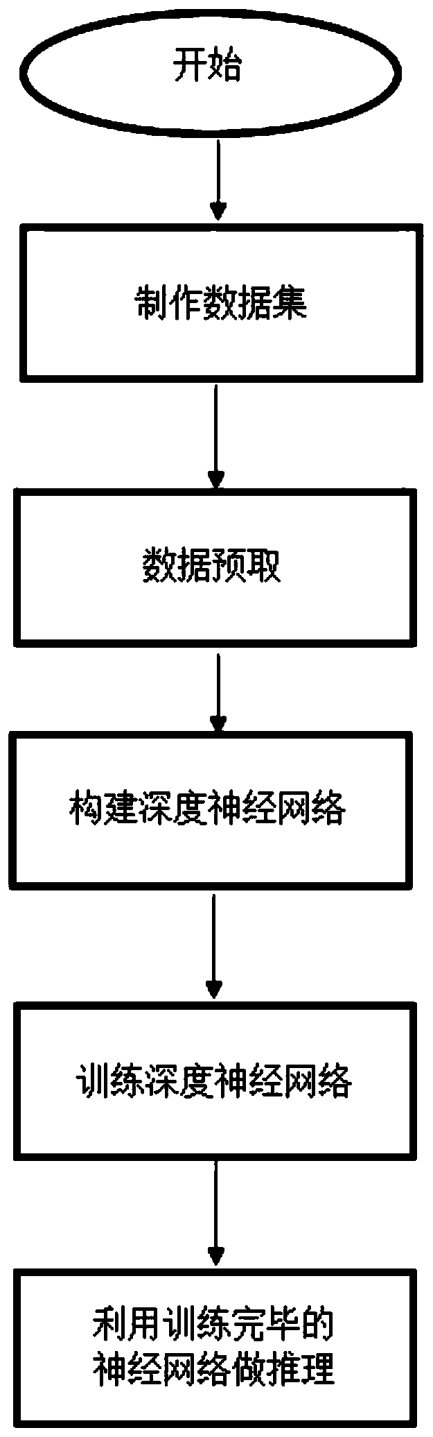
[0056] In the present embodiment, the method for automatically segmenting breast calcifications based on deep learning comprises the following steps:

[0057] (1) Make a data set

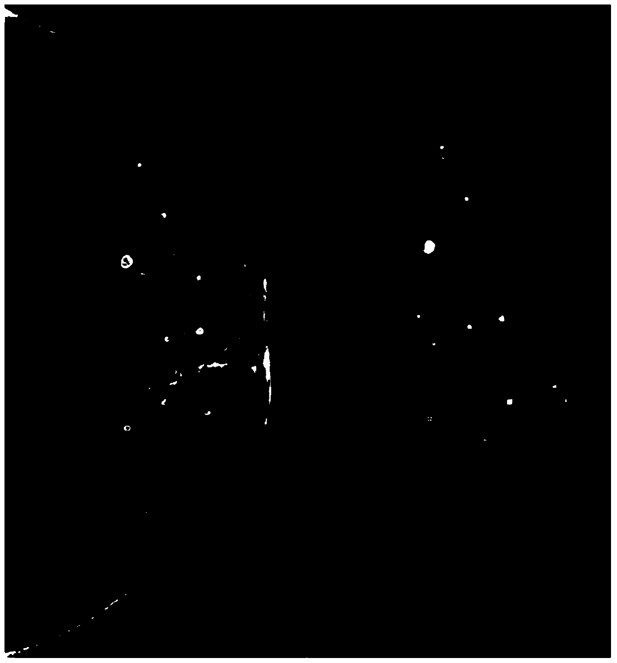
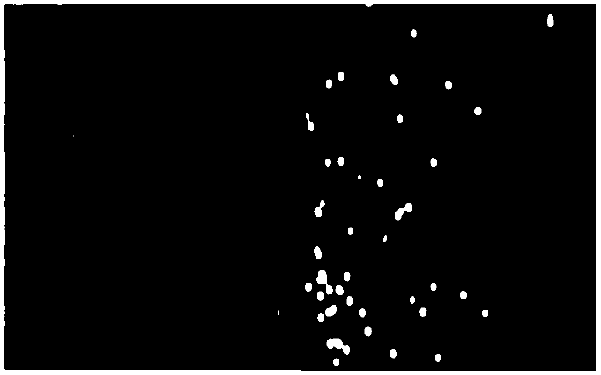
[0058] Desensitized breast X-ray images in DICOM format were obtained from no less than 10,000 medical records. Each case contained 4 images of left and right breast CC mid-axis and MLO lateral oblique views; at least 4 professional doctors read the images and performed mammograms. The radiograph calcification points are cross-labeled, and the images with consistent labels are used to construct the data set;

[0059] The difference between medical imaging and images in other professional fields is that the annotation of medical images requires the experience of professional doctors in film reading. Therefore, in this example, at least 4 professional doctors (su...


PUM


Abstract

The invention relates to a mammary gland X-ray auxiliary diagnosis technology, and aims to provide a method for automatically segmenting mammary gland calcification points based on deep learning. Themethod comprises the steps of making a data set; pre-fetching the data; constructing a deep convolutional neural network; training a deep convolutional neural network by using the normalized data set;and reasoning a to-be-detected image by using the trained deep convolutional neural network. According to the method, by introducing related technologies of deep convolutional neural network learningand training, all calcification points on the mammary gland X-ray image can be rapidly and automatically segmented. Based on the application of the invention, the accuracy of judging canceration by adoctor can be improved.

Description

technical field [0001] The invention relates to mammary gland X-ray assisted diagnosis technology, in particular to a method for automatically segmenting mammary gland calcification points based on deep learning. Background technique [0002] Computer-aided diagnosis is an important topic in the field of medical imaging. With the rise of artificial intelligence, computer image processing and pattern recognition have been more and more used in the field of assisted medical diagnosis. By obtaining different types of medical images, the lesions Segmentation and benign and malignant identification can assist doctors to observe the lesion more clearly and identify the characteristics of the lesion more accurately, which is of great significance. [0003] Breast cancer has developed into one of the most common cancers in women, and studies have shown that the cure rate for stage I breast cancer can reach 95%. Therefore, the earlier breast cancer is detected, the sooner interventi...

Claims


Application Information

Patent Timeline
Patent Type & Authority Applications(China)
IPC IPC(8): G06T7/11G06T7/00G06N3/04G06N3/08G16H30/40G16H50/20
CPCG06T7/11G06T7/0012G06N3/08G16H30/40G16H50/20G06T2207/10116G06T2207/20081G06T2207/20084G06T2207/30068G06N3/045
Inventor 吴法张宁子李康安
Owner ZHEJIANG DE IMAGE SOLUTIONS CO LTD
Why Eureka
Try Eureka