Cell detection and identification system and method

A technology for identifying systems and cells, applied in the field of cell detection and identification systems, and can solve problems such as affecting identification accuracy

Pending Publication Date: 2020-02-07
WUHAN LANDING INTELLIGENCE MEDICAL CO LTD
View PDF8 Cites 2 Cited by
  • Summary
  • Abstract
  • Description
  • Claims
  • Application Information

AI Technical Summary

Problems solved by technology

In the above scheme, in the process of converting a color image to a grayscale image, more information is discarded, which affects the subsequent recognition accuracy

Method used


Image

  • Cell detection and identification system and method
  • Cell detection and identification system and method
  • Cell detection and identification system and method

Examples


Embodiment 1

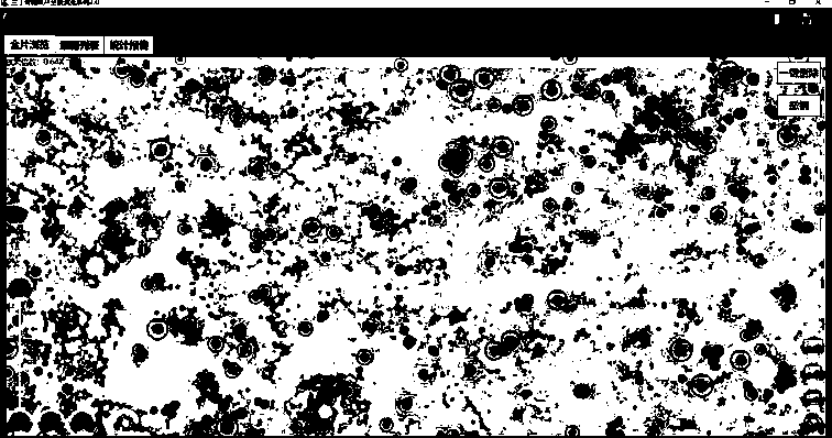
[0157] Such as Figure 1~6 Among them, a cell detection and identification system, which includes a microscopic scanning device, and the microscopic scanning device is used to obtain microscopic images;

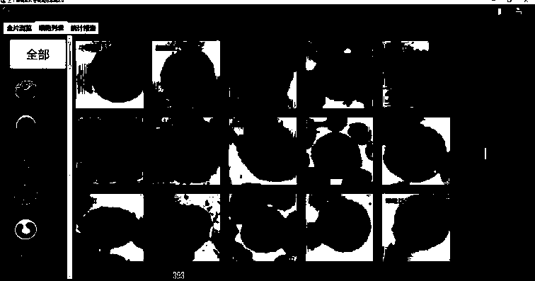
[0158] In the computer, multiple images of a single sample are stitched together, such as Figure 10a , 10b As shown in , extract according to the features of cell nuclei in the spliced ​​image to obtain a microscopic image of a single cell nuclei;

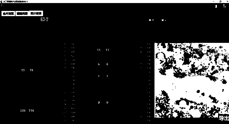
[0159] According to the labeled cells, the artificial intelligence program after model training is used to classify the microscopic images of single cell nuclei;

[0160] Such as Figure 11 As shown in , the artificial intelligence program preferably uses a convolutional neural network with a learning rate of 0.001. The number of result categories adopts num_classes = 3, corresponding to positive, negative and garbage respectively. Number of training rounds epochs = 300; image size: img_cols = 128 img_rows = 128; regularizat...

Embodiment 2

[0163]On the basis of Example 1, the preferred scheme is as Figure 3~6 In , the process of stitching images includes: field of view sub-block matching, field of view position fitting and block extraction;

[0164] Such as Figure 4 , Figure 10a As shown in , the process of visual subblock matching is:

[0165] Sa01, input, result set initialization M;

[0166] Sa02. Set the current field of view i as the first field of view;

[0167] Sa03. Find all adjacent visual field sets J of the current visual field i;

[0168] Sa04. Set the current adjacent field of view j as the first field of view in J;

[0169] Sa05. Find the possible overlapping areas Ri and Rj of the field of view i and field of view j;

[0170] Sa06. Rasterizing the template region Ri into a template sub-block set Pi;

[0171] Sa07. Arranging the template sub-block set Pi in descending order according to the dynamic range of the sub-blocks;

[0172] Sa08. Set the current template sub-block P as the first ...

Embodiment 3

[0217] On the basis of embodiment 1~2, preferred scheme is as figure 2 , Figure 7~9 In , the process of obtaining a microscopic image of a single nucleus is:

[0218] Sa100, detecting the characteristic points of the cell nucleus;

[0219] Reduce the image to multiple different ratios, preferably, the reduction ratios are: 0.3, 0.15, 0.08; extract feature points respectively;

[0220] Sa101. Preliminary screening, using the coordinates of the feature points to filter out the feature points that are too close to reduce the repeated extraction of cells; this step greatly improves the efficiency of recognition, especially the diagnosis efficiency of doctors.

[0221] In this example, if the distance of the feature points does not exceed half the radius of the cell, and half the radius is greater than 32, it is considered too close if the distance is less than 32 pixels, otherwise it is considered too close if it is less than half the cell radius. That is cell.Center.L1Distan...


PUM


Abstract

The invention provides a cell detection and identification system and method. The invention relates to the field of intelligent cell identification by using a computer, and discloses a cell identification system, which comprises a microscopic scanning device used for acquiring microscopic images. In a computer, multiple images of each sample are spliced in the computer, and extraction is made inspliced images according to cell nucleus characteristics to obtain a single cell nucleus microscopic image, According to marked cells, the single cell nucleus microscopic images are classified by using an artificial intelligence program after model training; and obtaining classified cell data based on the target. According to the invention, the identification accuracy and the identification efficiency can be greatly improved, especially the judgment time of doctors can be reduced, and the working efficiency is improved.

Description

technical field [0001] The invention relates to the field of intelligent identification of cells by using computers, in particular to a system and method for cell detection and identification. Background technique [0002] Cytological detection can quickly distinguish diseases according to the number and shape of cells, especially for the resolution and detection of tumor diseases. However, the efficiency of manual inspection in clinical practice is extremely low, and experienced doctors can only inspect about 50 samples per day. With the development of science and technology, automatic image analyzers have been used in cytopathological analysis. Can greatly improve efficiency. For example, the Chinese patent document CN 104881679 A describes a method for automatic detection of red blood cells in leucorrhea based on improved fuzzy recognition. The artificially found red blood cells are used to train the established neural network, and then the grayscale, binary According ...

Claims


Application Information

Patent Timeline
IPC IPC(8): G06T7/00G06T7/136G06T5/30
CPCG06T7/0014G06T7/136G06T5/30G06T2207/10056G06T2207/20036
Inventor 孙小蓉庞宝川肖笛
Owner WUHAN LANDING INTELLIGENCE MEDICAL CO LTD
Why Eureka
Try Eureka