
A method of ultrasonic super-resolution imaging

A super-resolution imaging and ultrasound technology, applied in ultrasonic/sonic/infrasonic diagnosis, sonic diagnosis, infrasonic diagnosis, etc., can solve the problems of increasing the concentration of microbubbles, limiting the imaging speed, and decreasing the speed of microbubbles, and reducing the system point diffusion function, suppressing scattering artifacts, reducing the effect of full width at half maximum

Active Publication Date: 2022-05-17
NANJING RAYGEN HEALTH CO LTD
View PDF13 Cites 0 Cited by
  • Summary
  • Abstract
  • Description
  • Claims
  • Application Information

AI Technical Summary

Problems solved by technology

However, due to the diffraction phenomenon when the size of the imaging object is smaller than the wavelength, the resolution of the imaging cannot break through the half-wavelength (clinical ultrasound is about a few hundred microns long), this phenomenon is called the diffraction limit
Although the contrast agent can greatly improve the signal-to-noise ratio of the imaging area, it still cannot overcome the diffraction limit, and it is difficult to see the topology of the overall blood flow from CEUS
[0003] However, ultrasonic super-resolution blood flow imaging based on microbubble localization has an irreconcilable contradiction between microbubble localization accuracy and imaging speed.
On the one hand, in order to ensure the accuracy of microbubble positioning, the concentration of microbubbles should not be too high, resulting in a decrease in the detection speed of microbubbles, thus limiting the imaging speed
On the other hand, increasing the concentration of microbubbles will lead to the sacrifice of localization accuracy, which will affect the quality of super-resolution imaging.
In addition, the concentration of microbubbles in blood vessels at various scales in the human body is not equal. Often, too low a concentration will make it difficult for microbubbles to perfuse into microvessels, while too high a concentration will make microbubbles no longer sparse in large blood streams.
Therefore, ultrasonic super-resolution blood flow imaging based on microbubble localization is difficult to balance accuracy and imaging speed.

Method used


Image

  • A method of ultrasonic super-resolution imaging
  • A method of ultrasonic super-resolution imaging
  • A method of ultrasonic super-resolution imaging

Examples


Embodiment Construction

[0015] The idea of ​​the present invention is that the following step diagrams and specific examples further illustrate the present invention in order to better understand the present invention, but the present invention is not limited to this specific example.

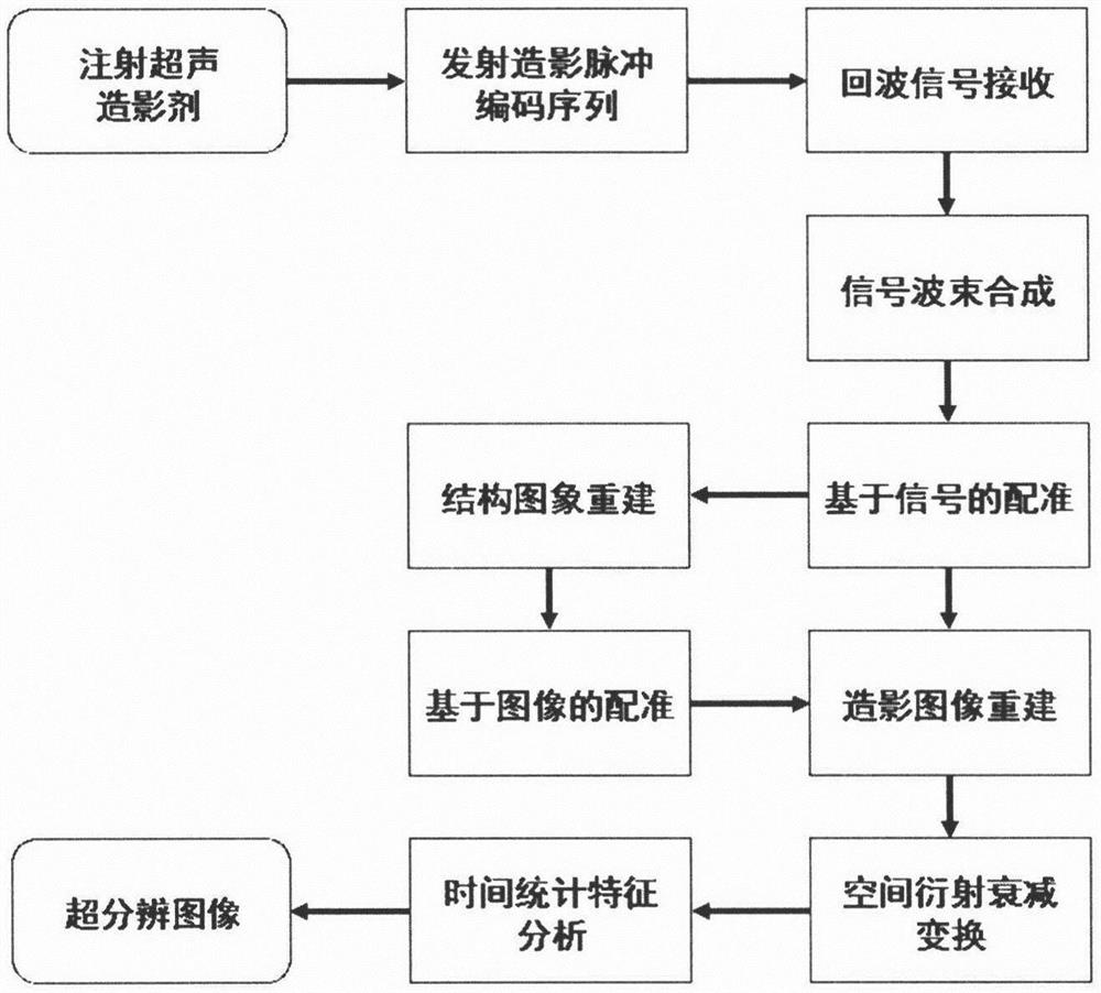
[0016] figure 1 is the flow chart of the present invention for reconstructing super-resolution images of blood flow, such as figure 1 Shown:

[0017] In step 1, the ultrasound contrast agent is firstly injected into the target to be imaged. The imaging area is detected with an ultrasound probe, and data acquisition begins when a contrast-enhanced signal appears. The contrast medium can be given as a one-time injection or as a continuous injection. A specific injection method for the biceps femoris of the lower limbs of Japanese big-eared white rabbits is to use 59 mg of sulfur hexafluoride microbubble freeze-dried powder for injection, Sonovi, dissolved in 5 mL of 0.9% sodium chloride solution to prepare a One-tim...


PUM


Abstract

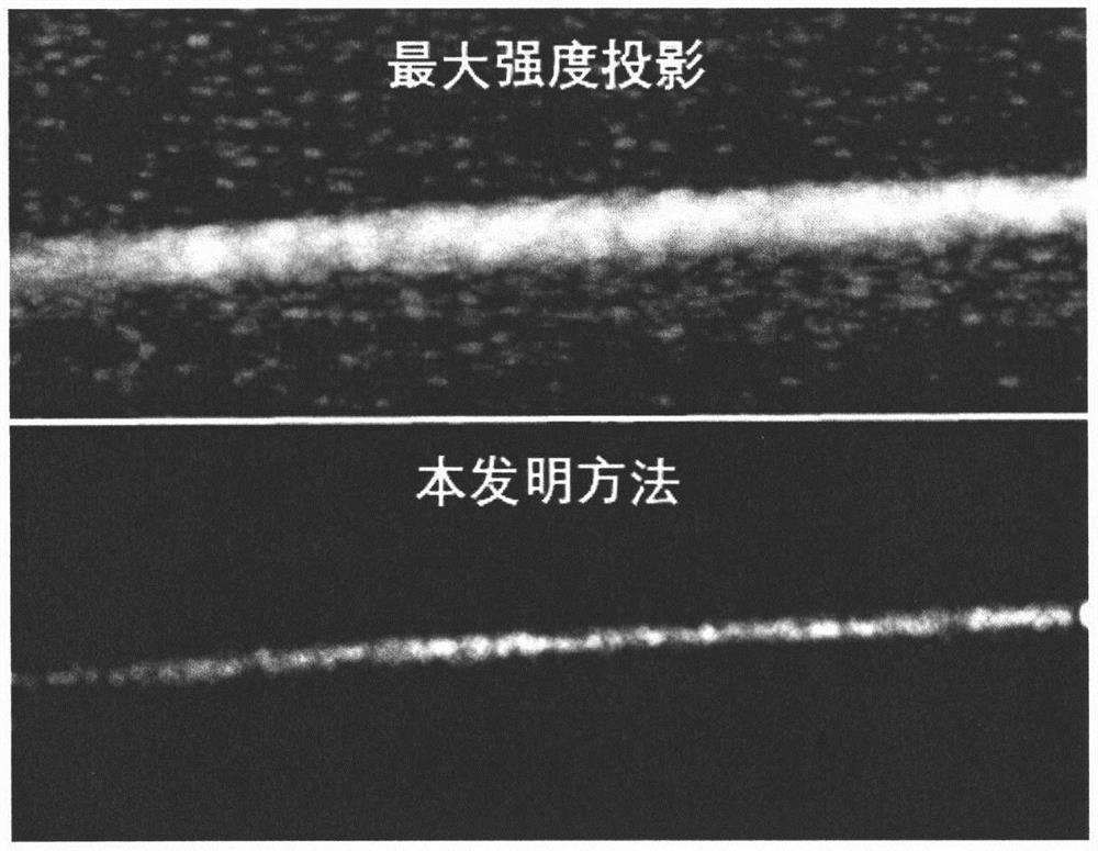
The invention discloses an ultrasonic super-resolution imaging method. This method first transmits space-time frequency coded ultrasound contrast pulse sequence to the region of interest injected with ultrasound contrast microbubbles to suppress the point spread function of the microbubble backscattering signal, and then performs spatial diffraction attenuation transformation or Position the center of the point spread function, converge the point spread function of the microbubble signal in the image, and finally perform time statistical analysis on the multi-frame converged image to enhance the signal of the center position of the microbubble, suppress background tissue and noise, and form a super-resolution image. This solution solves the difficult contradiction between microbubble positioning accuracy and imaging speed in ultrasonic super-resolution blood flow imaging, and achieves a substantial increase in super-resolution imaging rate.

Description

technical field [0001] The invention belongs to the field of ultrasonic imaging, in particular to an ultrasonic super-resolution imaging method. Background technique [0002] In the 1990s, the concept of ultrasound contrast agent entered the clinic for the first time. By injecting ultrasound contrast agent into the blood, the echo intensity of the area with blood flow can be enhanced, and the signal-to-noise ratio of the blood flow signal can be greatly improved. With the development of technology, the second-generation contrast agents represented by Optison and Sonovue entered the clinic, giving birth to contrast-enhanced ultrasound (CEUS), which can see the perfusion of blood flow (including small blood flow and slow blood flow) Condition. However, due to the diffraction phenomenon that occurs when the size of the imaging object is smaller than the wavelength, the resolution of the imaging cannot break through the half-wavelength (clinical ultrasound is about a few hundre...

Claims


Application Information

Patent Timeline
Patent Type & Authority Patents(China)
IPC IPC(8): A61B8/06
CPCA61B8/06A61B8/481A61B8/5215A61B8/5269
Inventor 张嘉宾
Owner NANJING RAYGEN HEALTH CO LTD
Learn More