
Multifunctional brain stereotaxic device

A stereotaxic and multi-functional technology, applied in the field of animal experiments, can solve the problems of labor cost and operation time, capillary rupture and bleeding, affecting positioning and drilling operation, etc., saving labor and time cost, drilling and micro volume The effect of precise injection and improved experimental validity

Pending Publication Date: 2020-02-14
TIANJIN KANGTING BIOLOGICAL ENG GRP CO LTD
View PDF0 Cites 1 Cited by
  • Summary
  • Abstract
  • Description
  • Claims
  • Application Information

AI Technical Summary

Problems solved by technology

[0004] 1. In the traditional stereotaxic instrument, there is no structure on the three-dimensional positioning rod that can lock the position of the scale. After the coordinate adjustment rod adjusts the coordinates, if you accidentally touch the adjustment rod, the coordinates of the adjustment rod will deviate, resulting in inaccurate positioning. re-locate;
[0005] 2. In the process of building an animal model with a traditional brain stereotaxic instrument, it is necessary to select different drill bits to position and drill holes according to the size of the selected animal head and the hardness of the animal skull, and to select micro-injectors of different specifications to inject the model into the positioning hole. To construct the required solution, it is necessary to replace drill bits and microinjectors of different types or specifications during actual operation. In traditional brain stereotaxic devices, when replacing drill bits and syringe needles, the original drill bits or syringes need to be disassembled and replaced with new ones, increasing Reduced labor costs and operating time
In the brain stereotaxic operation, when the skin on the top of the animal's head is opened for subsequent operations, the skin on both sides of the opening needs to be stretched to both sides, so as not to cover the skull and affect the positioning, punching and injection. The traditional brain stereotaxic device is not designed to support The device for opening the skin at the opening requires manual use of tweezers to stretch the skin at the opening to both sides to expose the skull. The skin is not fixed firmly with the tweezers, and the skin on both sides of the opening is easy to loosen, which affects the subsequent positioning and drilling operations;
[0007] 4. Because the animal's head is densely covered with capillaries, in the process of cutting the skin on the top of the animal's head to peel off the tissue, capillaries are often cut, resulting in capillary rupture and bleeding. In addition, various blood vessels are also distributed in the brain area below the animal's skull , When the drill bit is drilling at the coordinate point, the drill bit is also easy to touch the capillaries below the skull, causing the blood vessels in the positioning hole to rupture and bleed. After bleeding, the blood will cover the positioning hole, making the positioning hole blurred, affecting positioning and drilling;
[0008] 5. The traditional brain stereotaxic device does not have a collection and processing device for residual blood. After capillary bleeding in the head of the animal, it is necessary to manually clamp the hemostatic sponge or cotton ball to stop the bleeding. Hemostasis consumes labor costs and operating time, which affects the smooth progress of brain stereotaxic operation;
[0009] 6. During the stereotaxic process of animals, due to the individual differences of animals, various objective reasons such as the amount of anesthetics and the operator's technique may lead to the death of the animals, while the traditional stereotaxic devices are not designed for real-time monitoring of the animal's heartbeat status. The device cannot monitor the animal's heartbeat at any time, and cannot detect the signs of animal weakness in time and rescue it in time, resulting in the death of the animal and affecting the experimental operation.

Method used


Image

  • Multifunctional brain stereotaxic device
  • Multifunctional brain stereotaxic device
  • Multifunctional brain stereotaxic device

Examples


Embodiment Construction

[0030] The present invention will be further described in detail below in conjunction with the accompanying drawings and through specific embodiments. The following embodiments are only descriptive, not restrictive, and cannot limit the protection scope of the present invention.

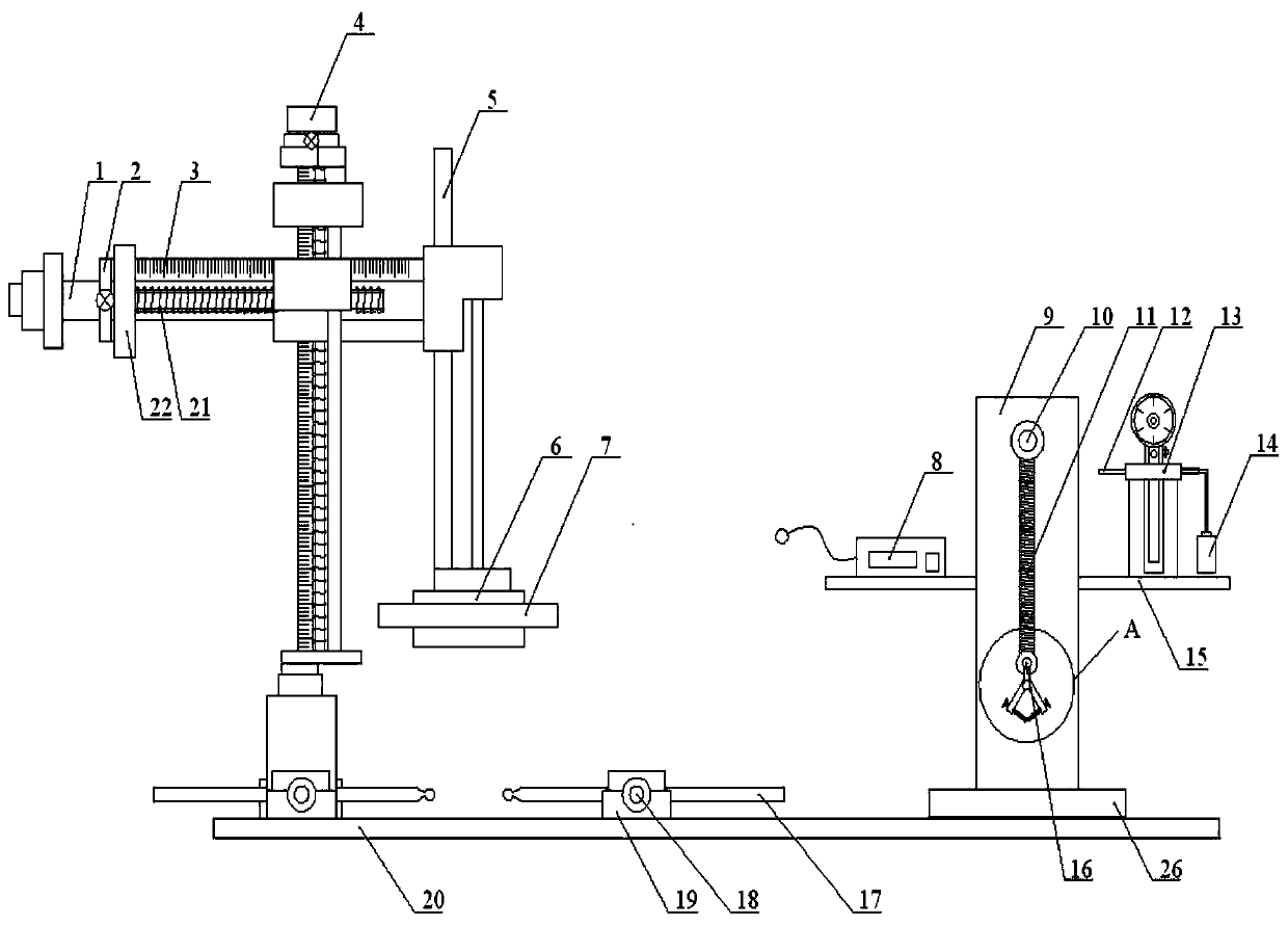
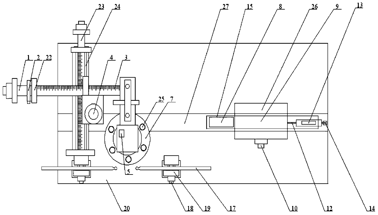
[0031] A multifunctional brain stereotaxic device, such as figure 1 , figure 2 As shown, it includes a fixing mechanism, a three-dimensional positioning mechanism and an auxiliary mechanism, wherein the fixing mechanism, the three-dimensional positioning mechanism and the auxiliary mechanism are all arranged on the bottom plate, and the three-dimensional positioning mechanism is arranged above the fixing mechanism. The slot 27, the auxiliary mechanism is slidably arranged in the transverse slide groove through the sliding block 26, and can slide to the top of the fixing mechanism and the bottom of the three-dimensional positioning mechanism.

[0032] Described fixing mechanism comprises base plate ...


PUM


Abstract

The invention relates to a multifunctional brain stereotaxic device. The device includes a fixing mechanism, a three-dimensional location mechanism and an auxiliary mechanism; the fixing mechanism, the three-dimensional location mechanism and the auxiliary mechanism are arranged on a base plate; the three-dimensional location mechanism is arranged above the fixing mechanism; the base plate is provided with a horizontal sliding groove; and the auxiliary mechanism is slidingly arranged in the horizontal sliding groove, and can slide to the upper part of the fixing mechanism and the lower part ofthe three-dimensional location mechanism. The device is scientific and reasonable in design and convenient in using, utilizes the fixing mechanism and the three-dimensional location mechanism to realize the fixing of an experimental rat, and can precisely and effectively perform adjusting and fixing on a three-dimensional coordinate; through the arrangement of a rotary limit disc, the device canbe suitable for drill bits and micro-syringes with various specifications, so that experiment accuracy and efficiency can be effectively enhanced; the supporting on the head skin of the experimental rat can be realized through an experimental supporting unit and an auxiliary unit of the auxiliary mechanism; through the auxiliary unit, the collection of residual blood and the monitoring on the heart of the experimental rat can be realized; and therefore, the integrated multifunctional brain stereotaxic device has high application values and good actual use effect.

Description

technical field [0001] The invention belongs to the field of animal experiments, and relates to a brain stereotaxic device, in particular to a multifunctional brain stereotaxic device. Background technique [0002] Brain stereotaxic apparatus, also known as brain fixation device (English name: stereotaxic apparatus), it uses the three-dimensional coordinate system stipulated by the marks on the outside of the skull or other reference points to determine the position of certain neural structures under the cortex, so that Directed stimulation, destruction, drug injection, guided potential and other researches were carried out under direct vision exposure. Animal brain stereotaxic apparatus is an important research equipment in the fields of neuroanatomy, neurophysiology, neuropharmacology, and neurosurgery. It is used for directional injection, stimulation, destruction, and guided potential operations on neural structures. It can be used for Parkinson's disease Establishment ...

Claims


Application Information

Patent Timeline
IPC IPC(8): A61D3/00A61B5/024
CPCA61B5/024A61B2503/40A61B2503/42A61D3/00A61D2003/003
Inventor 刘微微
Owner TIANJIN KANGTING BIOLOGICAL ENG GRP CO LTD
Learn More