Cartilage extractor for plastic surgery

An extractor and cartilage technology, applied in the field of medical devices, can solve the problems of increasing intercostal nerve and blood vessel damage, endangering the healthy life of the recipient, and puncturing the pleural cavity, so as to avoid vascular nerve damage and pleural cavity puncture. , the effect of simple operation

Pending Publication Date: 2020-02-21
WEST CHINA HOSPITAL SICHUAN UNIV
View PDF0 Cites 2 Cited by
  • Summary
  • Abstract
  • Description
  • Claims
  • Application Information

AI Technical Summary

Problems solved by technology

However, at present, most surgeons still use the traditional method of directly using rib hooks and scalpels to remove costal cartilage. This requires two-person cooperation, which wastes resources and time, and inc...

Method used


Image

  • Cartilage extractor for plastic surgery
  • Cartilage extractor for plastic surgery
  • Cartilage extractor for plastic surgery

Examples


Embodiment 1

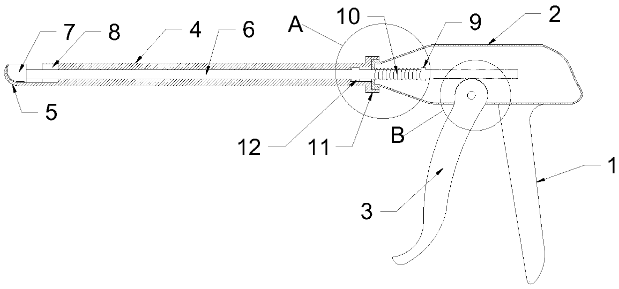
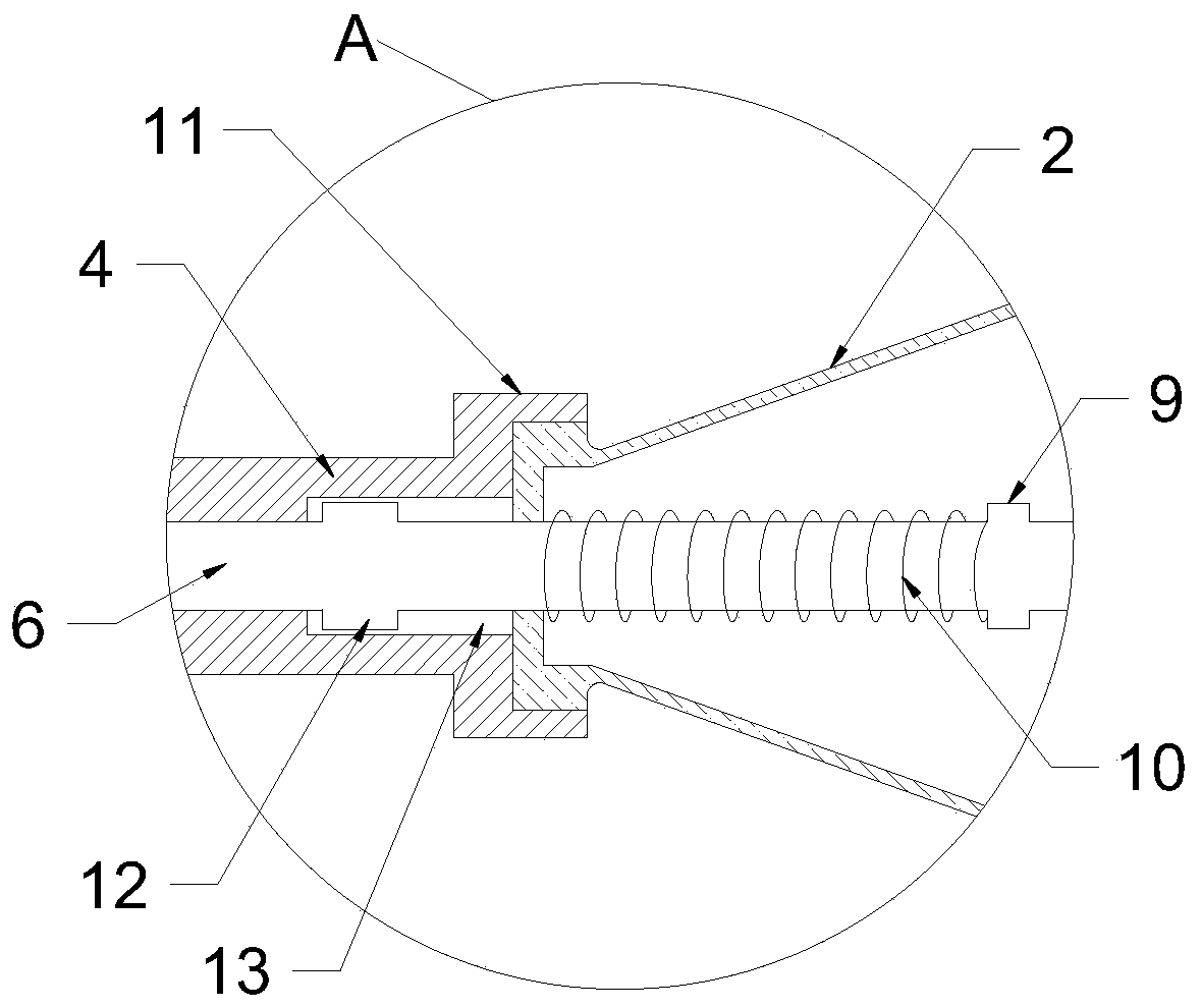
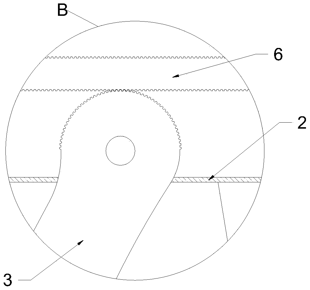
[0021] see Figure 1-4 , a cartilage extractor for plastic surgery, comprising a housing 2 with a handle 1, a firing rod 3 is hinged on the housing 2; A crotch 5 is provided, and the front end of the crotch 5 is an arc-shaped blunt metal sheet; a dowel 6 is inserted in the extension tube 4, and one end of the dowel 6 extends into the inner cavity of the housing 2, and the dowel The other end of 6 is equipped with a blade 7, the blade portion of the blade 7 can be attached to the inner wall of the crotch 5, and the front end of the extension tube 4 is provided with a storage groove 8 with a thickness greater than that of the blade 7; The 6 ends are provided with a plurality of tooth grooves evenly spaced, and the edge of the hinged end of the firing rod 3 is provided with teeth that can engage with the tooth grooves.

[0022] In this example, when the costal cartilage needs to be cut, a scalpel is used to make an incision on the side wall of the chest corresponding to the cost...

Embodiment 2

[0028] see Figure 1-6 , a cartilage extractor for plastic surgery, comprising a housing 2 with a handle 1, a firing rod 3 is hinged on the housing 2; A crotch 5 is provided, and the front end of the crotch 5 is an arc-shaped blunt metal sheet; a dowel 6 is inserted in the extension tube 4, and one end of the dowel 6 extends into the inner cavity of the housing 2, and the dowel The other end of 6 is equipped with a blade 7, the blade portion of the blade 7 can be attached to the inner wall of the crotch 5, and the front end of the extension tube 4 is provided with a storage groove 8 with a thickness greater than that of the blade 7; The 6 ends are provided with a plurality of tooth grooves evenly spaced, and the edge of the hinged end of the firing rod 3 is provided with teeth that can engage with the tooth grooves.

[0029] The inner wall of the crotch 5 is provided with a knife-edge groove 14 at the position corresponding to the blade of the blade 7. The setting of the knif...


PUM


Abstract

The invention relates to the technical field of medical instruments, and discloses a cartilage extractor for plastic surgery. The cartilage extractor comprises a shell with a handle, wherein a triggerrod is hinged on the shell; an extension pipe is arranged at the front end of the shell, a hook is arranged at one end, far away from the shell, of the extension pipe, and the front end of the hook is a round blunt metal sheet; a force transmission rod is inserted into the extension pipe, one end of the force transmission rod extends into the shell, and a blade is arranged at the other end of theforce transmission rod; a plurality of tooth sockets are arranged at the end of the force transmission rod positioned in an inner cavity of the shell; and an edge of the hinge end of the trigger rodis provided with teeth which can be meshed with the tooth sockets. According to the invention, a costal cartilage is hooked by the hook, and then the trigger rod controls a blade to cut the costal cartilage, so that the stripped costal cartilage can be hooked and cut. The whole process can be operated by one person, operation is simple, and operation time is shortened. In addition, the blade partof the blade can be attached to the inner wall of the hook, so that the costal cartilage can be completely cut at one time, and safety of a cutting process and integrity of the broken end of the costal cartilage are improved.

Description

technical field [0001] The invention relates to the technical field of medical devices, in particular to a cartilage extractor for plastic surgery. Background technique [0002] In recent years, with the development of plastic surgery technology and the improvement of people's aesthetic standards, as well as the influence of foreign plastic surgery colleagues, more and more doctors have begun to pay attention to the fine structure and shape of the tip of the nose. At present, in rhinoplasty, autologous costal cartilage combined with silica gel or puffy material for comprehensive rhinoplasty and nose shape adjustment has become the mainstream trend, especially for nasal tip shape reshaping. Autologous costal cartilage is recognized as the best material. Transplantation of costal cartilage can significantly change the shape and rotation of the nasal tip, and play a role in raising and lowering the nasal tip. However, at present, most surgeons still use the traditional method ...

Claims


Application Information

Patent Timeline
IPC IPC(8): A61B17/16
CPCA61B17/1693
Inventor 曹畅李棋孟晓彦张艳阁
Owner WEST CHINA HOSPITAL SICHUAN UNIV
Why Eureka
Try Eureka