Phase contrast microscopic imaging system and imaging method thereof

A micro-imaging and phase-contrast technology, which is applied in microscopes, image communications, color televisions, etc., can solve the problems of high thickness requirements, sample tissue observation that cannot be thick, and no application of human biopsy. Simple and easy-to-operate effects

Active Publication Date: 2020-03-27
ANKON MEDICAL TECH (SHANGHAI) CO LTD
View PDF6 Cites 0 Cited by
  • Summary
  • Abstract
  • Description
  • Claims
  • Application Information

AI Technical Summary

Problems solved by technology

Existing phase-contrast microscopy techniques can only observe slices with sample thicknesses in the order of microns, and have high requirements for thickness, and cannot observe thicker sample tissues (millimeters), and have no real significance in human biopsy. application

Method used


Image

  • Phase contrast microscopic imaging system and imaging method thereof
  • Phase contrast microscopic imaging system and imaging method thereof
  • Phase contrast microscopic imaging system and imaging method thereof

Examples


Embodiment Construction

[0028] The technical solutions in the embodiments of the present invention will be clearly and completely described below in conjunction with the accompanying drawings in the embodiments of the present invention. Obviously, the described embodiments are only some of the embodiments of the present invention, not all of them. . Based on the embodiments of the present invention, all other embodiments obtained by persons of ordinary skill in the art without making creative efforts belong to the protection scope of the present invention.

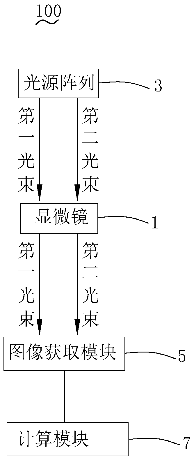
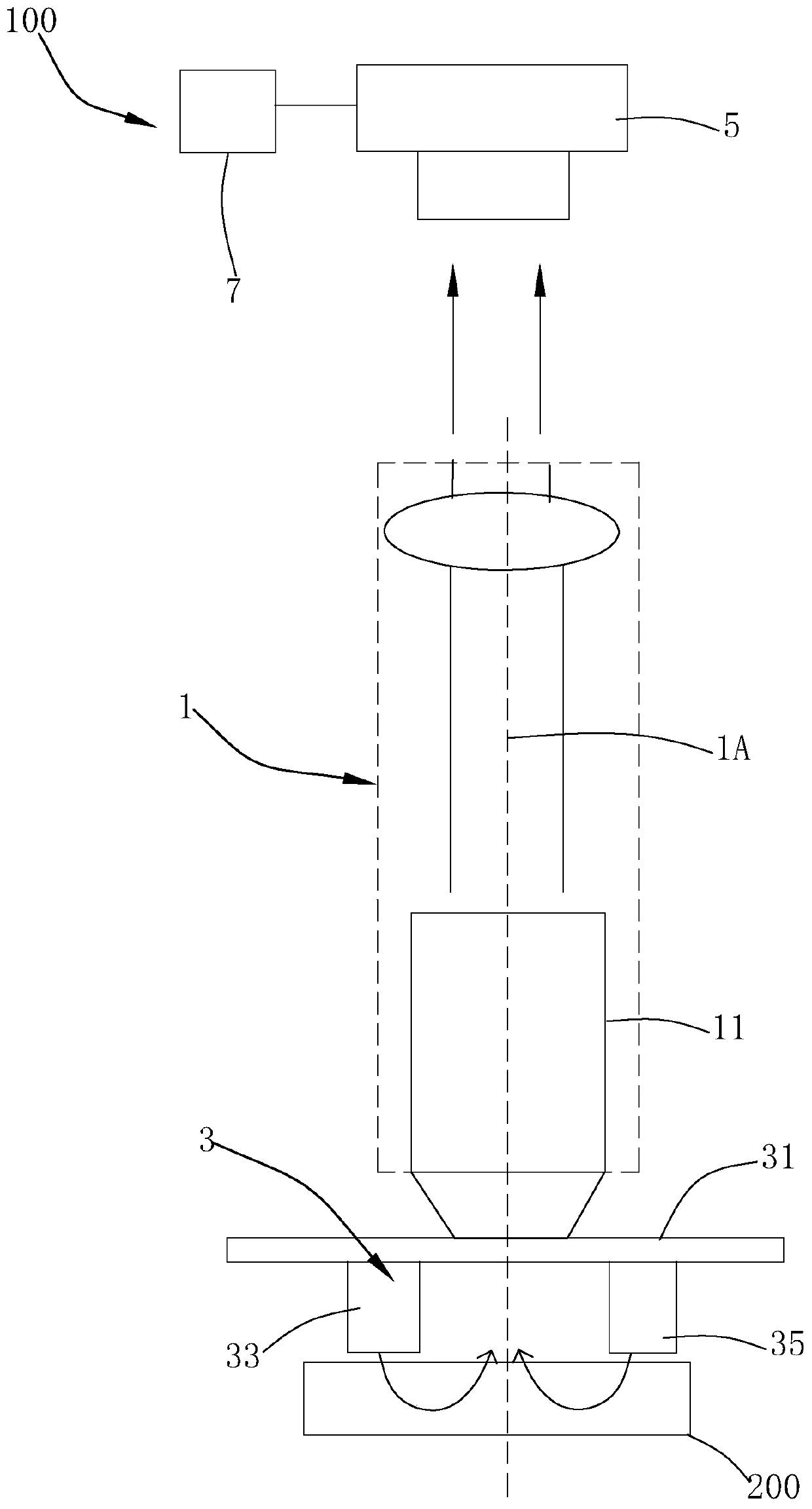
[0029] Please refer to Figure 1 to Figure 3 , the phase contrast microscopy imaging system 100 includes a microscope 1 with an optical axis 1A, a light source array 3 , an image acquisition module 5 and a computing module 7 .

[0030] The microscope 1 includes an objective lens 11 .

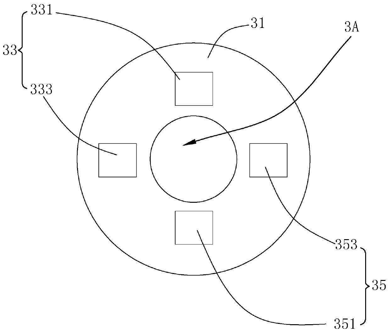
[0031] The light source array 3 includes a fixture 31 fixed on the object side of the objective lens 11 and having a through hole 3A, and two fixtures 31 fixed o...


PUM


Abstract

The invention provides a phase contrast microscopic imaging system with a simple structure. The phase contrast microscopic imaging system comprises a microscope with an optical axis, a light source array, an image acquisition module and a calculation module; the microscope includes an objective lens; the light source array comprises a fixture with a through hole and two light source groups arranged around the through hole, the two light source groups emit a first light beam and a second light beam which are symmetrically arranged relative to the optical axis so as to irradiate a sample, and the light beams enter the sample, scatter, obliquely penetrate through the sample, are transmitted out and are collected by a microscope after passing through the through hole; the image acquisition module acquires a first image corresponding to the first light beam and a second image corresponding to the second light beam on the image side of the microscope; the calculation module is used for recovering the primary color component of the first image and the primary color component of the second image and subtracting the primary color component obtained by the first image from the primary colorcomponent obtained by the second image to obtain a phase contrast image. The imaging method of the phase contrast microscopic imaging system is simple and easy to operate.

Description

【Technical field】 [0001] The invention relates to the technical field of microscopic imaging, in particular to a phase contrast microscopic imaging system and an imaging method thereof. 【Background technique】 [0002] The development of microscopic imaging technology has brought a whole new world to the human eye. People can see hundreds of "new" tiny animals and plants, as well as the internal structure of everything from human bodies to plant fibers. , help scientists discover new species, and help doctors treat diseases. [0003] Phase-contrast microscopy is used to visualize unstained specimens. As the light wave passes through living cells and undyed biological specimens, its wavelength and amplitude do not change, which cannot be observed by the human eye, but its phase changes. Phase contrast microscopy changes the phase difference and uses light diffraction and interference phenomena to change the phase difference into an amplitude difference to observe living cell...

Claims


Application Information

Patent Timeline
Patent Type & Authority Applications(China)
IPC IPC(8): G02B21/06G02B21/36G01N21/84
CPCG02B21/06G02B21/361G01N21/84G02B21/14G02B21/367G02B21/082H04N9/645
Inventor 段晓东王青青游庆虎
Owner ANKON MEDICAL TECH (SHANGHAI) CO LTD
Why Eureka
Try Eureka