Back-portable visual in-vivo calcium imaging device

An imaging device and portable technology, which is applied in medical science, veterinary instruments, sensors, etc., can solve the problems that the imaging device cannot fix the focal length, cannot capture stable calcium signals, and is not very mature, so as to achieve abundant calcium signal recording methods , not easy to move, not easy to relax

Pending Publication Date: 2020-04-10
FOURTH MILITARY MEDICAL UNIVERSITY
View PDF0 Cites 1 Cited by
  • Summary
  • Abstract
  • Description
  • Claims
  • Application Information

AI Technical Summary

Problems solved by technology

However, based on the characteristics of the anatomical structure and physiological functions of the spine, the application and research of the in vivo calcium imaging technology of the spinal cord is not very mature at present, because the spine itself is a movable joint with physiological curvature, and mice can easily turn when they move freely. The head bites off the imaging device fixed on the back, and because the spine keeps swinging with the movement, the imaging device will not be able to fix the focus, so that stable calcium signals cannot be taken. perfect stage

Method used


Image

  • Back-portable visual in-vivo calcium imaging device
  • Back-portable visual in-vivo calcium imaging device
  • Back-portable visual in-vivo calcium imaging device

Examples


Embodiment Construction

[0017] In order to make the object, technical solution and advantages of the present invention clearer, the implementation manner of the present invention will be further described in detail below in conjunction with the accompanying drawings.

[0018] It should be noted that when a component is considered to be "set on" another component, it can be directly set on another component, or there may be a central component at the same time; when a component is considered to be "connected" to another component, It can be directly connected to another component, or there may be an intermediate component at the same time.

[0019] Unless otherwise defined, all technical and scientific terms used in the present invention have the same meaning as commonly understood by one of ordinary skill in the technical field of the present invention. The terms used in the description of the present invention are only for the purpose of describing specific embodiments, and are not intended to limit...


PUM


Abstract

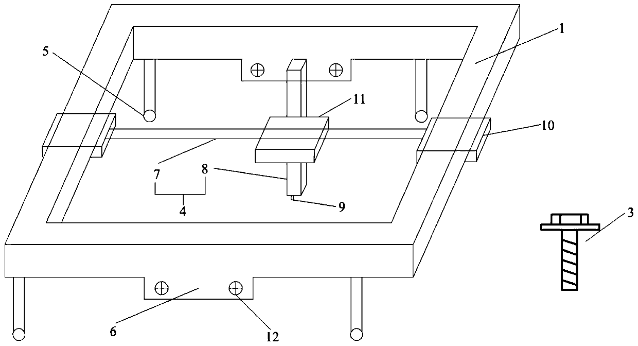
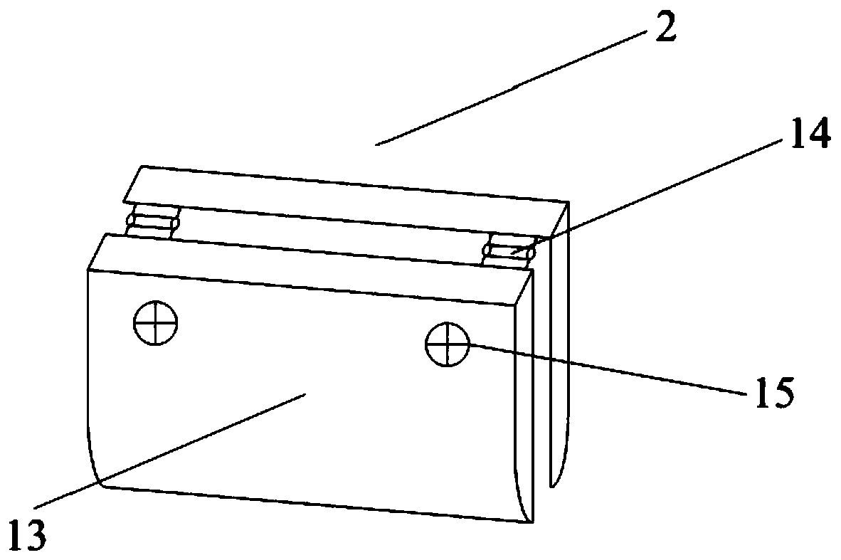
The invention discloses a back-portable visual in-vivo calcium imaging device, and belongs to the technical field of in-vivo calcium imaging. The device includes a device body, a spine holder and fixing screws; the device body includes a track pulley system, four universal wheels and a spine holder base, the track pulley system is composed of two mutually perpendicular metal rods, wherein the twomutually perpendicular metal rods include a horizontal track and a longitudinal track, and the bottom end of the longitudinal track is provided with a microscope fixing platform; and the spine holderis connected to the spine holder base of the device main body through the four fixing screws, and is located at the inner lower part of the device main body. According to the mouse back-portable in-vivo calcium imaging device provided by the invention, a mouse spine can be fixed and is not easy to move, the mouse spine can be combined with the device main body carrying an ultramicroscope, and thedevice has the advantages of clever design, a simple and reasonable structure, stable clamping, good flexibility, convenient use, and strong compatibility, and can make calcium signal recording methods more abundant.

Description

technical field [0001] The invention relates to the technical field of in vivo calcium imaging, in particular to a back-carried visualized in vivo calcium imaging device. Background technique [0002] Calcium imaging refers to the method of using calcium ion indicator to monitor the calcium ion concentration in tissues. In the study of the nervous system, in vivo or in vitro experiments, calcium imaging technology is widely used to monitor the changes of calcium ions in hundreds or thousands of neurons at the same time, so as to detect the activity of neurons Condition. As soon as this technology appeared, it was sought after by neuroscientists all over the world, and it is still the most direct means for people to observe neuron activity. [0003] In recent years, with the development of optogenetics and in vivo calcium imaging technology, people have a very complete application method for brain in vivo calcium imaging technology. However, based on the characteristics of...

Claims


Application Information

Patent Timeline
Patent Type & Authority Applications(China)
IPC IPC(8): A61B5/00A61D3/00
CPCA61B5/0077A61B5/0082A61B5/407A61B5/6802A61B2503/40A61B2503/42A61D3/00
Inventor 王福东罗层解柔刚褚文广吴文斌崔雅婷从翔宇田志成
Owner FOURTH MILITARY MEDICAL UNIVERSITY
Why Eureka
Try Eureka