End-to-end mammary gland ultrasound image segmentation method based on Distance-AttU-Net (Distance-AttU-Net)

A technology of ultrasound images and mammary glands, applied in the fields of deep learning, computer vision and medical images, can solve problems such as low precision and complex data processing steps, and achieve high segmentation accuracy, good scalability and applicability

Pending Publication Date: 2020-04-21
BEIJING UNIV OF TECH
View PDF3 Cites 2 Cited by
  • Summary
  • Abstract
  • Description
  • Claims
  • Application Information

AI Technical Summary

Problems solved by technology

[0007] In order to solve the problem of low accuracy and complicated data processing steps in the segmentation technology of existing breast ultrasound images when dealing with multi-size lesions

Method used


Image

  • End-to-end mammary gland ultrasound image segmentation method based on Distance-AttU-Net (Distance-AttU-Net)
  • End-to-end mammary gland ultrasound image segmentation method based on Distance-AttU-Net (Distance-AttU-Net)
  • End-to-end mammary gland ultrasound image segmentation method based on Distance-AttU-Net (Distance-AttU-Net)

Examples


Embodiment Construction

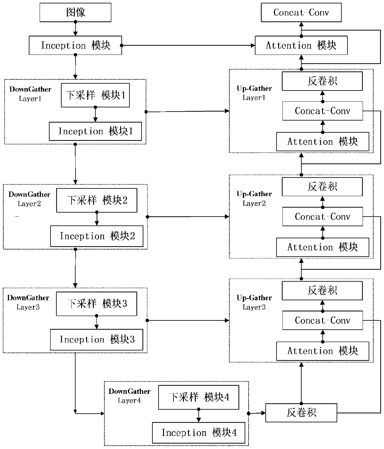
[0029] In order to make the object, technical solution and advantages of the present invention clearer, the present invention will be further described in detail below in conjunction with specific embodiments and with reference to the accompanying drawings.

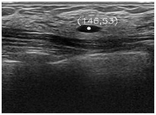
[0030] The calculation process of the distance from the pixel point to the center point of the lesion area: the calculation result is as follows figure 2 shown.

[0031] Step S1, distance calculation:

[0032] Step S1.1, using the public opencv library to calculate the coordinates of the lesion area. Since the real labeled image is the lesion area segmented on the original breast ultrasound image, the center point is directly calculated on the lesion area in the real labeled image. First, use the findContours function to find the lesion area, and then use the moments function to calculate the central moment of the area found in the previous step;

[0033] Step S1.2, record the center coordinates of the lesion area obt...


PUM


Abstract

The invention discloses an end-to-end mammary gland ultrasonic image segmentation method based on Distance-AttU-Net. The method comprises the following steps: firstly, an original mammary gland ultrasonic image is directly used as input of a network, a trained loss value is calculated by using a self-defined loss function, and a network model is trained by optimizing the loss value and updating model parameters through back propagation until the model converges. Through the training of the network, the network itself has the capability of distinguishing the lesion from the similar regions around the lesion, and finally the lesion region in the mammary gland ultrasonic image is segmented. Through testing on the public data set B, the network model has high segmentation capability, it is proved that the mammary gland lesion segmentation model based on the ultrasonic image with excellent performance can be trained under the condition that training samples are few, and the mammary gland lesion segmentation model based on the ultrasonic image has the advantages of end-to-end segmentation, multi-scale lesion processing, high discrimination and the like.

Description

technical field [0001] The invention relates to the fields of deep learning, computer vision and medical images, in particular to an end-to-end breast ultrasound image segmentation method based on Distance-AttU-Net. Background technique [0002] There are many ways to diagnose breast cancer. Ultrasound imaging has become the most effective way to diagnose breast lesions due to its advantages of no radiation, low cost, and fast imaging. However, the judgment and understanding of breast ultrasound images requires rich clinical experience of doctors, and the workload of interpretation is relatively large. Relying entirely on manual processing by doctors is prone to fatigue, leading to a further increase in misdiagnosis rates. Therefore, in order to reduce the burden on doctors and improve diagnostic results, it is extremely important to study reliable methods for intelligent segmentation of ultrasound breast lesions. [0003] At present, the methods for ultrasonic breast image...

Claims


Application Information

Patent Timeline
Patent Type & Authority Applications(China)
IPC IPC(8): G06T7/11G06T7/194
CPCG06T7/11G06T7/194G06T2207/10132G06T2207/20081G06T2207/20084G06T2207/30068
Inventor 杨新武侯海娥
Owner BEIJING UNIV OF TECH
Why Eureka
Try Eureka