Kidney segmentation method in CT image based on deep learning

A CT image and deep learning technology, applied in the field of kidney segmentation in CT images based on deep learning, can solve problems such as confusion of kidney and spleen, and achieve the effect of eliminating interference and fast convergence.

Active Publication Date: 2020-05-05
北京小白世纪网络科技有限公司
View PDF2 Cites 1 Cited by
  • Summary
  • Abstract
  • Description
  • Claims
  • Application Information

AI Technical Summary

Problems solved by technology

[0005] For example, the neural network Unet does have a certain effect on segmentation, but for images with similar shapes and similar CT values, such as figure 1

Method used


Image

  • Kidney segmentation method in CT image based on deep learning
  • Kidney segmentation method in CT image based on deep learning
  • Kidney segmentation method in CT image based on deep learning

Examples


Embodiment Construction

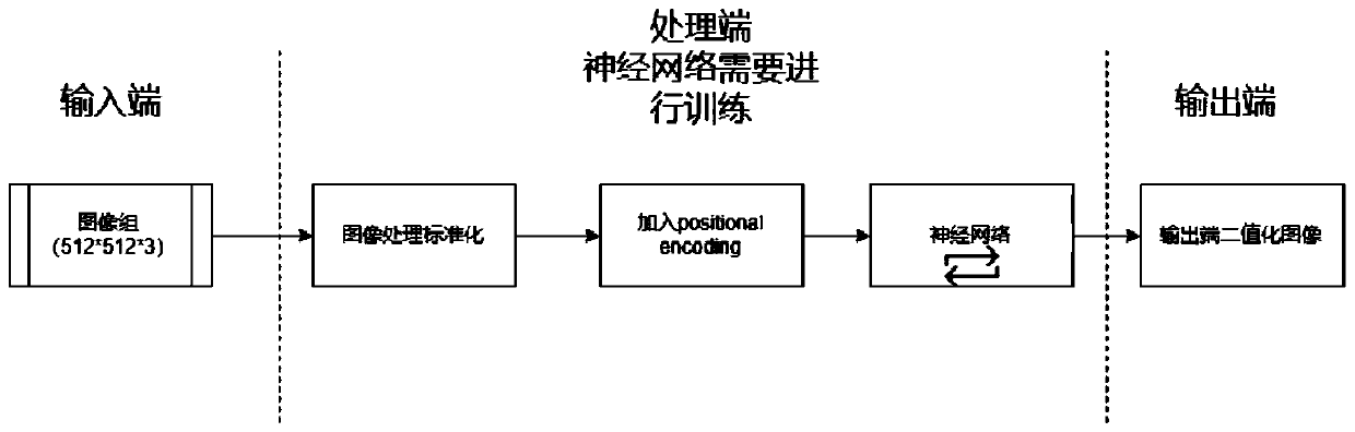
[0036] The present invention provides a kidney CT image segmentation network method that adds position coding and attention mechanism, allowing the neural network to segment the kidney without being confused with the spleen or other organs through back-propagation. The present invention will be described in detail below in conjunction with the accompanying drawings.

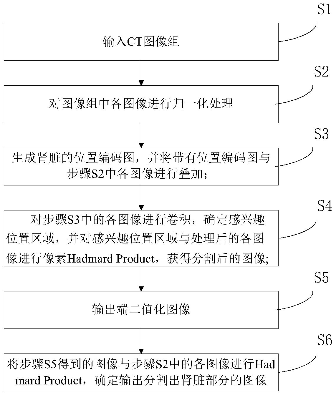
[0037] Such as Figure 2-3 As shown, the present invention provides a method for segmenting kidneys in CT images based on deep learning, comprising the following steps:

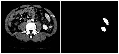
[0038] S1. Input the CT image group; in this embodiment, the CT image group can be 3 consecutive CT images, the length and width are 512*512, and the pixel value range is -1000~2000HU. During training, multiple groups of images can be imported for training. The three consecutive images have the information of the upper and lower layers, so that the model can predict the mask of the middle layer through the information of the upper and lower lay...


PUM


Abstract

The invention discloses a kidney segmentation method in a CT image based on deep learning. The method comprises the following steps: inputting a CT image group; carrying out normalization processing on each image in the image group; generating a position coding graph of the kidney, and superposing the position coding graph with each image; carrying out convolution on all the images, determining aninterested position area, carrying out pixel Hadamard code product on all the processed images, and obtaining segmented images; binarizing the image at the output end; and carrying out Hadamard codeproduct on the obtained image and each image after normalization processing, and determining and outputting the image of which the kidney part is segmented. According to the method, the position codesare added when the neural network is used for training the images, so that the problem that the spleen and the kidney are difficult to distinguish can be well distinguished and solved; besides, an attention mechanism is used, so that the network fitting convergence speed is higher, the network can pay attention to the kidney position area, and therefore, spleen interference is eliminated.

Description

technical field [0001] The invention relates to the technical field of image segmentation, in particular to a method for segmenting kidneys in CT images based on deep learning. Background technique [0002] With the large-scale growth of image data on the Internet, image segmentation technology has been widely concerned and applied. Especially in the segmentation of the medical field, the cost of segmentation for doctors is high, the efficiency of segmentation is low, and the segmentation standards are uneven. [0003] Existing medical segmentation technology is generally based on traditional computer vision technology, project CT value (-1000Hu, 1000Hu) to RGB space (0-255), manually extract features, use heuristic algorithm according to doctor's experience, use the extracted Features such as dimensionality reduction and machine learning algorithms. Such a method has many advantages. For example, the experience of doctors can be well automated. For example, a series of ma...

Claims


Application Information

Patent Timeline
IPC IPC(8): G06T7/11G06N3/08G06N3/04
CPCG06T7/11G06N3/084G06T2207/10081G06T2207/20084G06T2207/20081G06T2207/30084G06N3/045
Inventor 杜强李剑楠郭雨晨聂方兴张兴
Owner 北京小白世纪网络科技有限公司
Why Eureka
Try Eureka