
Device for endoscopic optoacoustic imaging, in particular for endoscopic optoacoustic imaging of cavities and hollow objects

A technology of photoacoustic imaging and endoscopy, which can solve the problems of lack of molecular contrast, etc.

Pending Publication Date: 2020-06-02
健康与环境慕尼黑德国研究中心赫姆霍茨中心(有限公司)
View PDF8 Cites 1 Cited by
  • Summary
  • Abstract
  • Description
  • Claims
  • Application Information

AI Technical Summary

Problems solved by technology

However, OCT has the disadvantage of an inherent lack of molecular contrast (the ability to distinguish the molecule of interest from other molecules)

Method used


Image

  • Device for endoscopic optoacoustic imaging, in particular for endoscopic optoacoustic imaging of cavities and hollow objects
  • Device for endoscopic optoacoustic imaging, in particular for endoscopic optoacoustic imaging of cavities and hollow objects
  • Device for endoscopic optoacoustic imaging, in particular for endoscopic optoacoustic imaging of cavities and hollow objects

Examples


Embodiment Construction

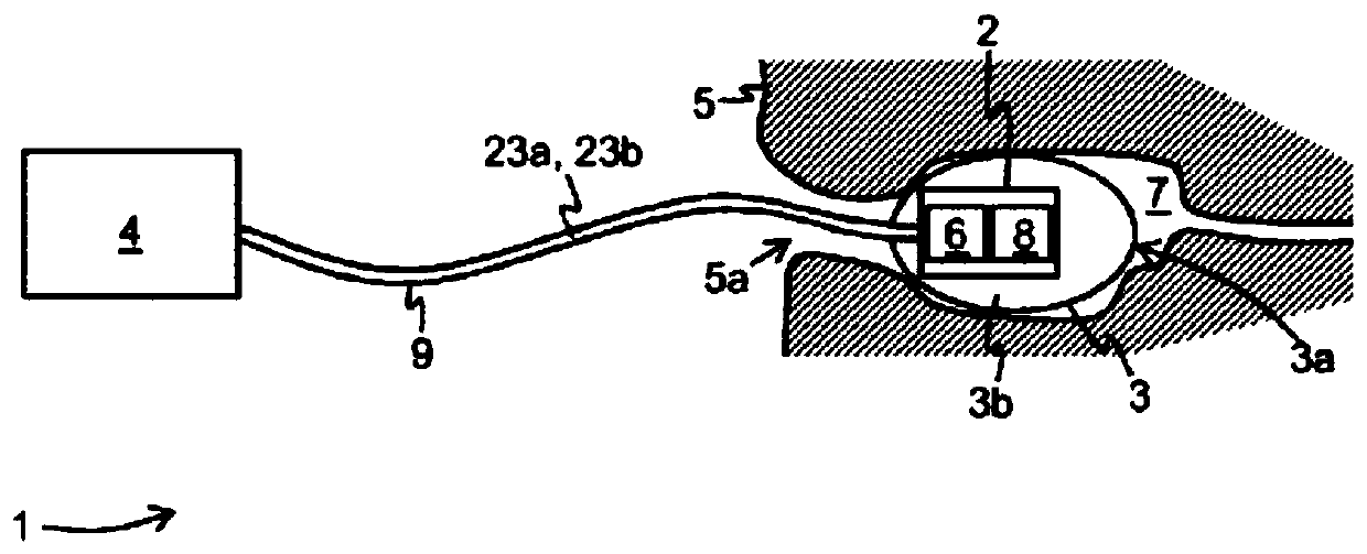
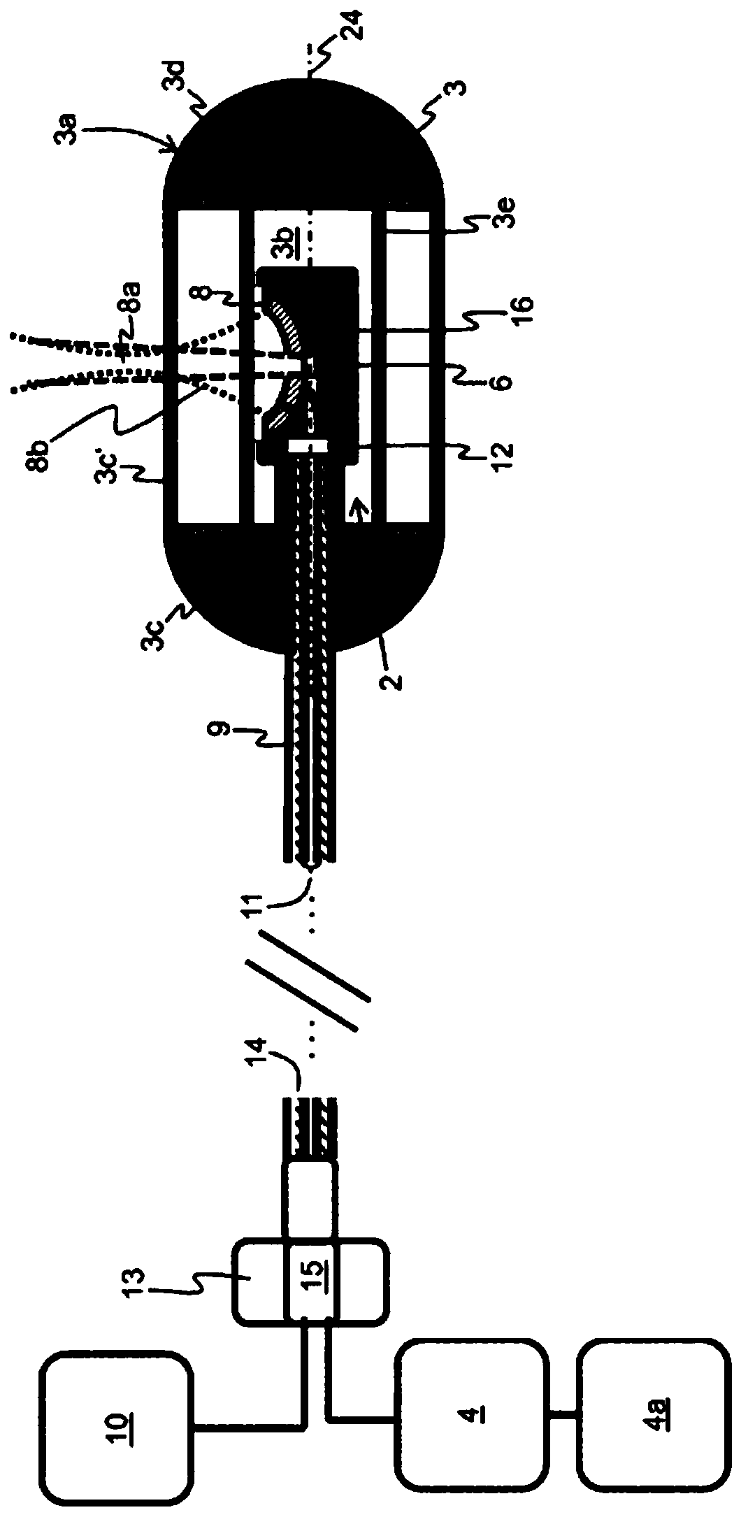
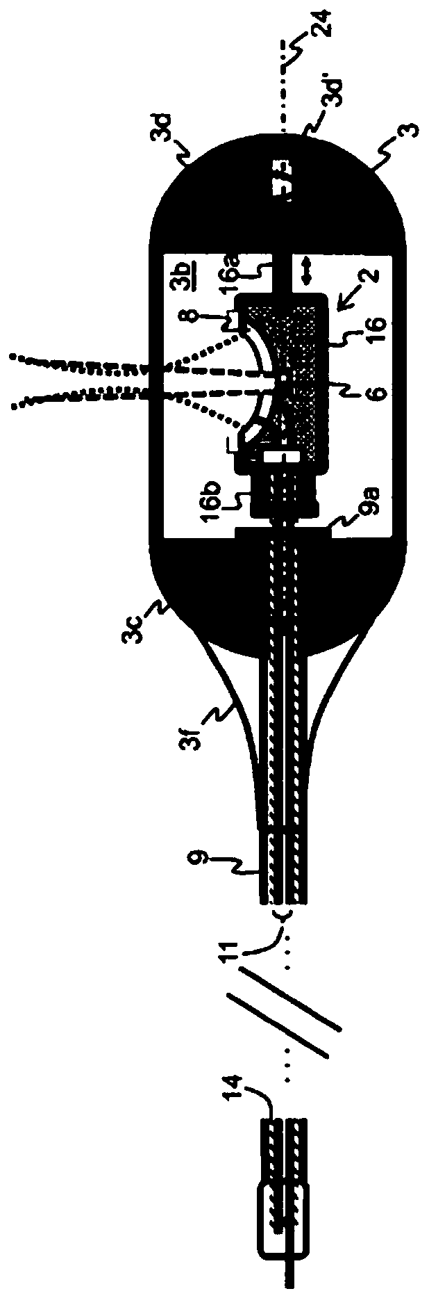
[0075] figure 1 An example of a device 1 for endoscopic photoacoustic imaging comprising an imaging unit 2 , a position stabilization structure 3 and a processing unit 4 is shown. In the following, the position stabilizing structure 3 is also referred to as "position stabilizing unit".

[0076] The imaging unit 2 is configured to be inserted into an object 5, such as the gastrointestinal (GI) tract of a human body, via a natural orifice 5a of the human body, and includes an illumination unit 6 configured to emit electromagnetic radiation, particularly at wavelengths in the visible or infrared Light in the spectral range is used to illuminate the region of interest 7 (eg the cavity wall within the object 5). Acoustic waves generated in the region of interest 7 in response to irradiation with electromagnetic radiation are detected by the detection unit 8, whereby corresponding detection signals are generated.

[0077] The imaging unit 2 is connected to the processing unit 4 vi...


PUM


Abstract

The invention relates to a device (1) for endoscopic optoacoustic imaging comprising an imaging unit (2) configured to be at least partially inserted into an object (5), said imaging unit (2) comprising an irradiation unit (6) configured to irradiate a region of interest (7) inside the object (5) with electromagnetic radiation and a detection unit (8) comprising at least one ultrasound transducerconfigured to detect ultrasound waves generated in the region of interest (7) in response to irradiating the region of interest (7) with the electromagnetic radiation and to generate according detection signals, wherein the at least one ultrasound transducer exhibits a field of view (8b) being at least partially located in the irradiated region of interest (7). A position stabilizing structure (3)with an outer face (3a) and an interior (3b) is configured to stabilize and / or fix the imaging unit (2) in a position and / or orientation in the object (5) by bringing the outer face (3a) of the position stabilizing structure (3) into contact with the object (5). The device (1) further comprises a processing unit (4) configured to generate an optoacoustic image (23b) of the region of interest (7)based on the detection signals.

Description

technical field [0001] The invention relates to a device for endoscopic photoacoustic imaging, in particular for endoscopic photoacoustic imaging of cavities and hollow objects, according to the independent claims. Background technique [0002] Endoscopy is a standard technique for examination of the gastrointestinal (GI) tract and is based on flexible imaging probes, such as optical video endoscopes inserted through natural lumens (e.g., in the digestive system) for visual observations of interest. cavity organs. However, detection of early-stage disease lacking specificity and staging ability by visual perception is often not possible. This is also limited by the surface structure. Disease-related features that manifest beneath the tissue surface, such as lesions that do not develop into cancer and possible invasion of the gastrointestinal wall by cancer or metastatic invasion of adjacent lymph nodes, are thus not revealed. For diagnostic purposes, a gastroenterologist ...

Claims


Application Information

Patent Timeline
IPC IPC(8): A61B5/00A61B1/04A61B8/12
CPCA61B5/6861A61B1/041A61B5/0066A61B5/0068A61B5/0071A61B5/0075A61B5/0095A61B8/12A61B1/00165A61B1/00148A61B1/00147
Inventor 瓦西利斯·恩齐亚克里斯托斯乔治·塞尔吉亚迪斯克里斯蒂安·扎基安安德烈亚斯·布勒
Owner 健康与环境慕尼黑德国研究中心赫姆霍茨中心(有限公司)
Learn More