
Cervical cell image classification method based on graph convolutional neural network

A convolutional neural network and cervical cell technology, applied in the field of digital image processing, can solve the problems of pathologists’ burden and fatigue of film reading, achieve high classification accuracy, improve diagnostic efficiency and accuracy, broad application value and market prospect

Active Publication Date: 2020-06-12
HEFEI UNIV OF TECH
View PDF5 Cites 15 Cited by
  • Summary
  • Abstract
  • Description
  • Claims
  • Application Information

AI Technical Summary

Problems solved by technology

A cervical cell smear usually contains tens of thousands of cervical cells, and the screening process brings a great burden to pathologists, and the phenomenon of reading fatigue occurs from time to time

Method used


Image

  • Cervical cell image classification method based on graph convolutional neural network
  • Cervical cell image classification method based on graph convolutional neural network
  • Cervical cell image classification method based on graph convolutional neural network

Examples


Embodiment Construction

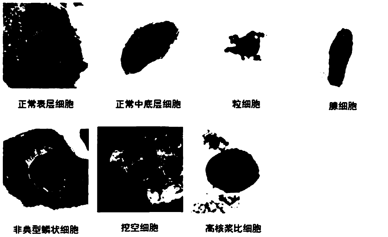
[0030] Such as figure 1 As shown, step 1: prepare training samples; classify the cervical cell images marked by doctors to obtain seven types of samples, the seven types of samples include normal superficial cells, normal middle and bottom cells, granulocytes, gland cells (cervical tube cells), SARS Type squamous cells, koilocytes, high nuclear plasma ratio cells; such as figure 2 shown;

[0031] Step 2: Obtain the 1024-dimensional feature representation of the training sample; remove the last fully connected layer of the pre-trained dense convolutional neural network to obtain a feature extractor, and input each training sample into the feature extractor to obtain a 1024-dimensional feature vector , which is used as the feature representation of the sample.

[0032] Step 3: Construct a sample feature relationship graph; use each class of training samples as a node of the relationship graph. Obtain the mean value of each sample feature as the feature of the node. Then cal...


PUM


Abstract

The invention discloses a cervical cell image classification method based on a graph convolutional neural network. Firstly, a cervix uteri cell image is prepared as a training sample; 1024-dimensionalfeature representation of a sample is acquired, a sample feature relation graph is constructed, a deep network based on a graph convolutional network is constructed, the sample and the sample featurerelation graph are sent to the deep network model for training, training is stopped after iteration is performed for a certain number of times, and network weight parameters are stored. When the method is used, a target image is segmented into a to-be-predicted area with cell nucleuses, then weight parameters and a network structure obtained through training are loaded, the to-be-predicted area is input into the to-be-predicted area, and a classification result can be obtained through calculation. According to the method, the cervical cell diagnosis accuracy and efficiency are improved, the diagnosis process of a pathologist is optimized, and the workload of the pathologist is reduced.

Description

technical field [0001] The present invention relates to the technical field of digital image processing, in particular to digital image classification technology, in particular to a method for cervical cell image classification based on a graph convolutional neural network. Background technique [0002] Cervical cell classification has a vital clinical application value in the early screening of cervical cancer. A cervical cell smear usually contains tens of thousands of cervical cells. The screening process brings a great burden to pathologists, and the phenomenon of film reading fatigue occurs from time to time. Therefore, a digital cervical cell classification method is needed to assist pathologists in the classification of cervical cells, reduce the burden of film reading for pathologists, and improve the diagnostic accuracy of pathologists. [0003] Graph Convolutional Networks (GCN) is a network structure that can perform convolution operations on data that exists in ...

Claims


Application Information

Patent Timeline
Patent Type & Authority Applications(China)
IPC IPC(8): G06K9/00G06K9/62G06N3/04G06N3/08
CPCG06N3/084G06V20/695G06V20/698G06N3/045G06F18/24
Inventor 史骏王若宇李俊代杰
Owner HEFEI UNIV OF TECH
Learn More