Device for high tibial osteotomy

A technology for osteotomy and tibial bone, which is applied in the field of high tibial osteotomy devices, which can solve the problems of shaking of the osteotomy end, cumbersome surgical steps, and large surgical trauma, so as to shorten the operation time, reduce the operation risk, and simplify the operation The effect of steps

Pending Publication Date: 2020-06-26
XIANGYA HOSPITAL CENT SOUTH UNIV
View PDF4 Cites 4 Cited by
  • Summary
  • Abstract
  • Description
  • Claims
  • Application Information

AI Technical Summary

Problems solved by technology

[0005] (1) For the "osteotomy" step, the selection of the osteotomy line depends on experience, and the intraoperative operations such as the direction of osteotomy and the depth of osteotomy are purely based on hand feeling, which is imprecise and easy to damage the surrounding tissues;
[0006] (2) For the step of "correcting the alignment and temporary fixation", correcting the alignment and angle of the tibia after osteotomy depends entirely on the operator's experience and intraoperative fluoroscopy confirmation, which requires multiple attempts and a large amount of intraoperative radiation exposure. Still inaccurate, often incomplete orthopedics;
[0009] (1) These devices generally can only complete the two steps of "assisting osteotomy" and "assisting temporary fixation to correct the line of force";
[0010] (2) The bone veneers of these devices are all concentrated in the

Method used


Image

  • Device for high tibial osteotomy
  • Device for high tibial osteotomy
  • Device for high tibial osteotomy

Examples


Embodiment Construction

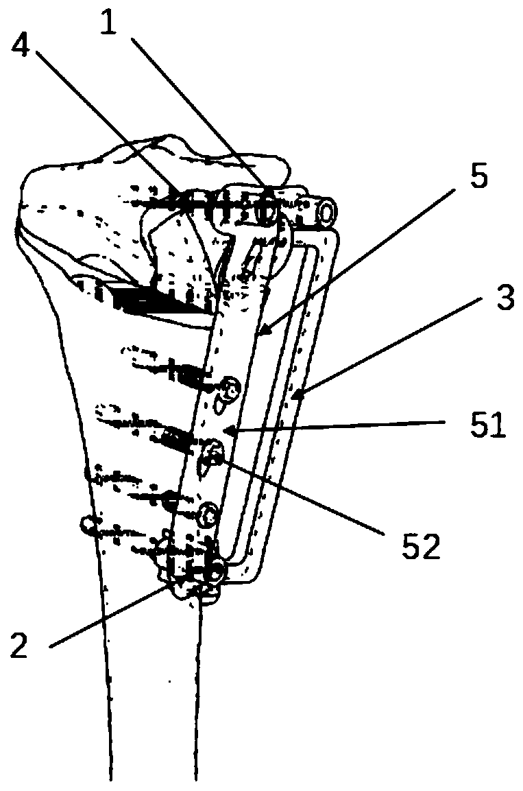
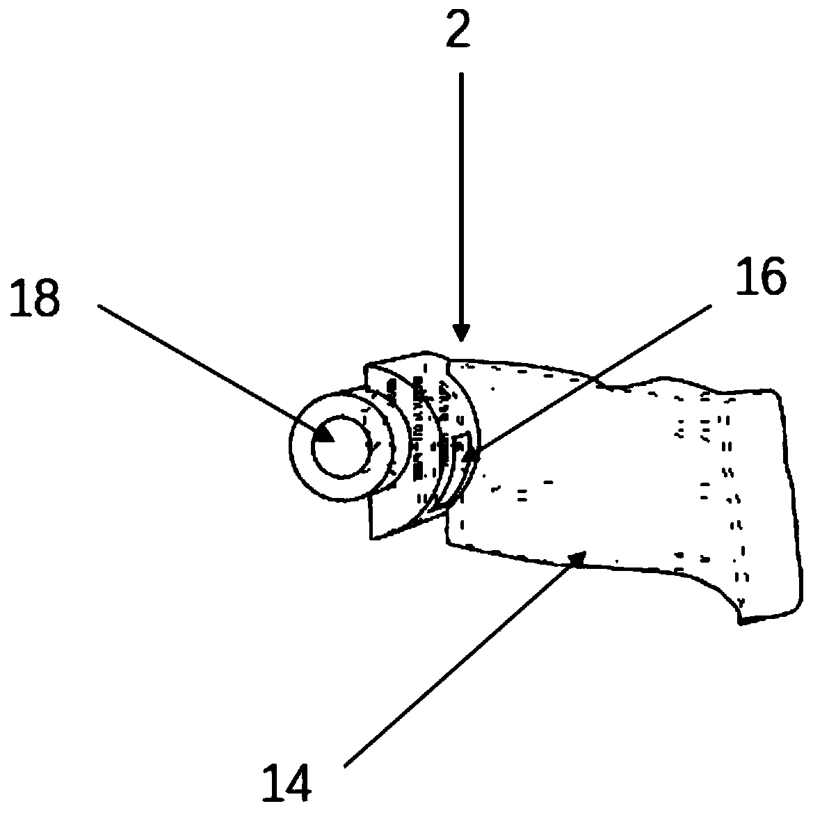
[0045] The technical solution of the present invention will be further described in detail below in conjunction with the accompanying drawings of the description: a high tibial osteotomy device, such as figure 1As shown, it includes a proximal part 1, a distal part 2, a bridge part 3, a Kirschner wire 4, a fixing part 5, and a calibration force line part 6; the proximal part 1 and the distal part 2 are located on two sides of the bridge part 3 The proximal and distal parts are fixed on the tibia through the Kirschner wire 4; the calibration force line part 6 is connected to the proximal part 1 through a pin, and the fixing part 5 is connected to the proximal and distal ends of the tibia along the 4 holes of the Kirschner wire. The fixing part 5 fits on the surface of the tibia after the line of force is corrected.

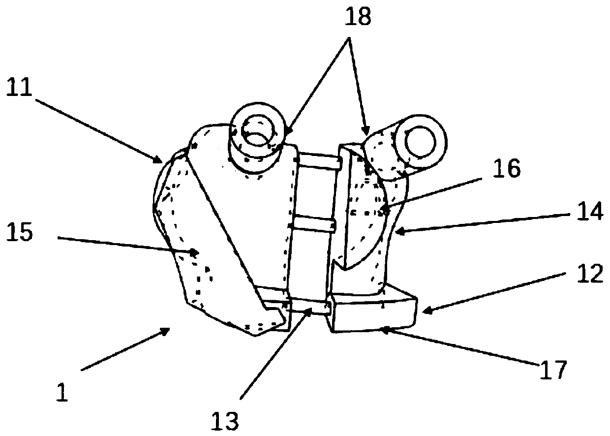
[0046] Further, such as figure 2 The proximal part 1 shown is divided into two parts, the longitudinal osteotomy area 11 and the transverse osteotomy area 12, th...


PUM


Abstract

The invention relates to the field of medical instruments, in particular to a device for high tibial osteotomy. The device comprises a near-end component, a far-end component, a connecting bridge component, Kirschner wires, a fixing component and a calibration force line component, wherein the near-end component and the far-end component are located at the two ends of the connecting bridge component respectively and fixed to a tibia through Kirschner wires; the calibration force line component is connected with the near-end component through a bolt; and the fixing component is attached to thesurface of the tibia having undergone force line correction and connected with the near end and the far end of the tibia through Kirschner wire hole sites. According to the invention, the design of bony wainscots for the near end and the far end of the tibia is adopted, so the area of a tibia incision near an osteotomy line can be reduced, an operative wound is narrowed, and tissue around the osteotomy line is better protected; the device integrates three functions of osteotomy, force line correction and temporary immobilization and steel plate installation in a high tibial osteotomy operation; operation steps are simplified, operation time is shortened, operation risks are reduced, and the effect of treating knee osteoarthritis through the high tibial osteotomy operation can be better promoted.

Description

technical field [0001] The invention relates to the field of medical instruments, in particular to a high tibial osteotomy device. Background technique [0002] When osteoarthritis occurs in the knee joint, the proximal end of the tibia (high position) is often deformed. After the deformation, the knee joint is deformed such as varus, which makes the force on the knee joint unbalanced, and then the symptoms of knee osteoarthritis appear or aggravate. [0003] Therefore, there is a clinical surgical method that can specifically treat knee arthritis in the above situation, that is, high tibial osteotomy. The doctor performs osteotomy on the proximal tibia of the patient, then restores the bone angle of the tibia, and restores the line of force of the lower limbs, and then uses a steel plate Screws are used for internal fixation, so that the force on the knee joint is balanced, so as to achieve the purpose of treating knee arthritis. The operation generally requires three step...

Claims


Application Information

Patent Timeline
IPC IPC(8): A61B34/10A61B17/90A61B17/80A61B17/56A61B17/17A61B17/16
CPCA61B17/1657A61B17/1675A61B17/1732A61B17/1764A61B17/56A61B17/8061A61B17/88A61B2017/564A61B34/10A61B2034/104A61B2034/105A61B2034/108A61B17/90
Inventor 钟达王成功雷鹏飞
Owner XIANGYA HOSPITAL CENT SOUTH UNIV
Why Eureka
Try Eureka