Cardiovascular image recognition system and method based on whole-heart seven-dimensional model

An image recognition and cardiovascular technology, applied in neural learning methods, biological neural network models, medical images, etc., can solve the problems of high subjective influence of cardiovascular imaging and inaccurate interpretation results, and improve the efficiency and accuracy of image acquisition. , strong compatibility and universality, the effect of ensuring accuracy

Active Publication Date: 2020-06-26
SICHUAN UNIV
View PDF6 Cites 4 Cited by
  • Summary
  • Abstract
  • Description
  • Claims
  • Application Information

AI Technical Summary

Problems solved by technology

[0009] In view of the above-mentioned deficiencies in the prior art, the cardiovascular image recognition system and method based on the seven-dimensional model of the whole heart provi...

Method used


Image

  • Cardiovascular image recognition system and method based on whole-heart seven-dimensional model
  • Cardiovascular image recognition system and method based on whole-heart seven-dimensional model
  • Cardiovascular image recognition system and method based on whole-heart seven-dimensional model

Examples


Embodiment 1

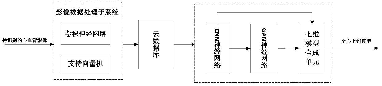
[0060] Such as figure 1 As shown, the cardiovascular image recognition system based on the whole-heart seven-dimensional model includes the sequentially connected image data processing subsystem, cloud database and whole-heart seven-dimensional model construction subsystem;

[0061] The image data processing subsystem is used to process the input cardiovascular images to be identified, accurately mark them, and upload them to the cloud database;

[0062] The cloud database is used to store all accurately labeled cardiovascular images;

[0063] The whole-heart seven-dimensional model construction subsystem is used to construct the corresponding whole-heart seven-dimensional model according to the required cardiovascular images in the cloud database, and realize the recognition of the cardiovascular images according to the constructed whole-heart seven-dimensional model.

[0064] The image data processing subsystem in the embodiment of the present invention is a convolutional n...

Embodiment 2

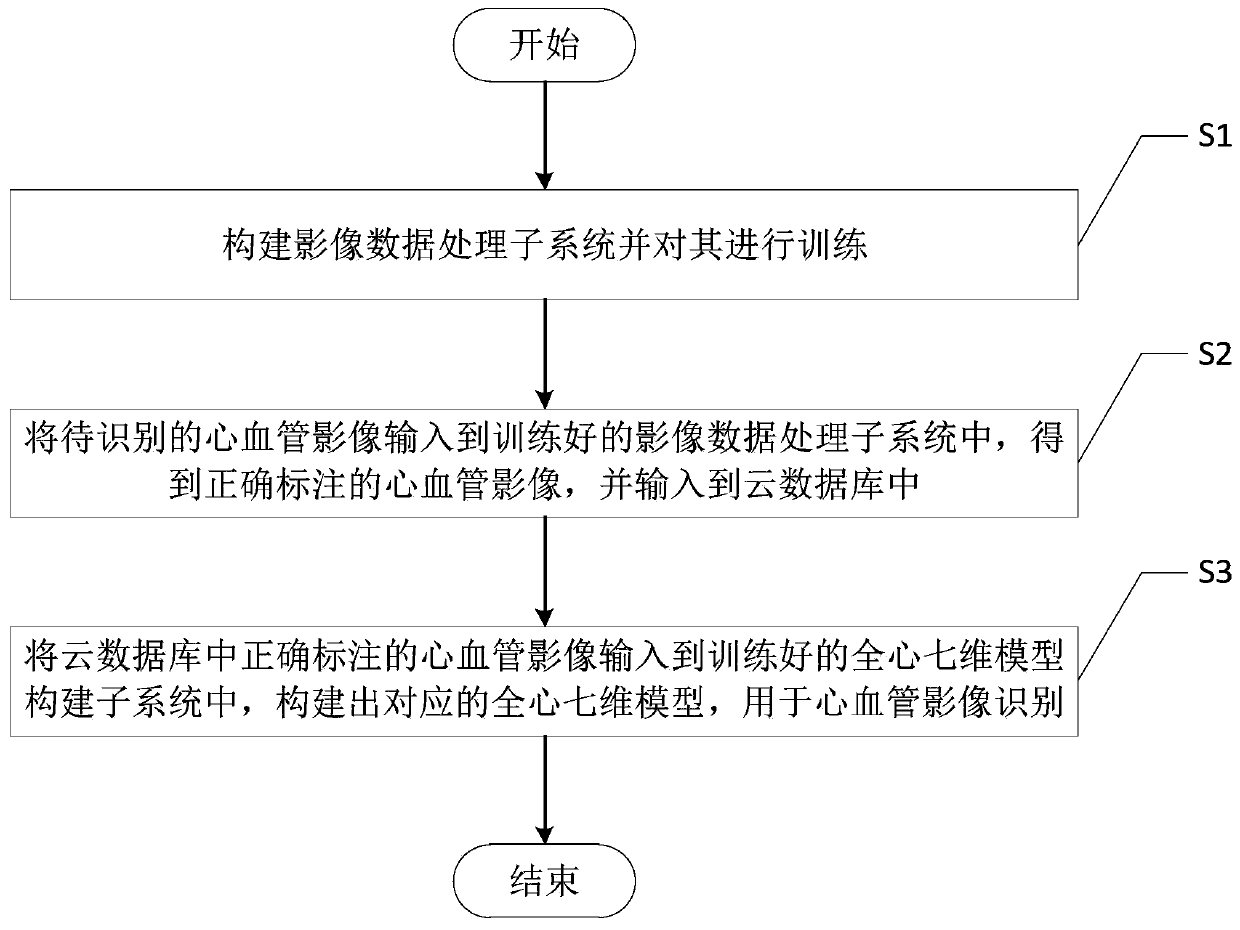
[0068] Such as figure 2 As shown, corresponding to the above-mentioned embodiment 1, the present invention also provides a cardiovascular image recognition method based on the whole-heart seven-dimensional model, comprising the following steps:

[0069] S1. Construct the image data processing subsystem and train it;

[0070] S2. Input the cardiovascular image to be identified into the trained image data processing subsystem, obtain the correctly marked cardiovascular image, and input it into the cloud database;

[0071] S3. Input the correctly labeled cardiovascular images in the cloud database into the trained whole-heart seven-dimensional model building subsystem, and construct a corresponding whole-heart seven-dimensional model for cardiovascular image recognition.

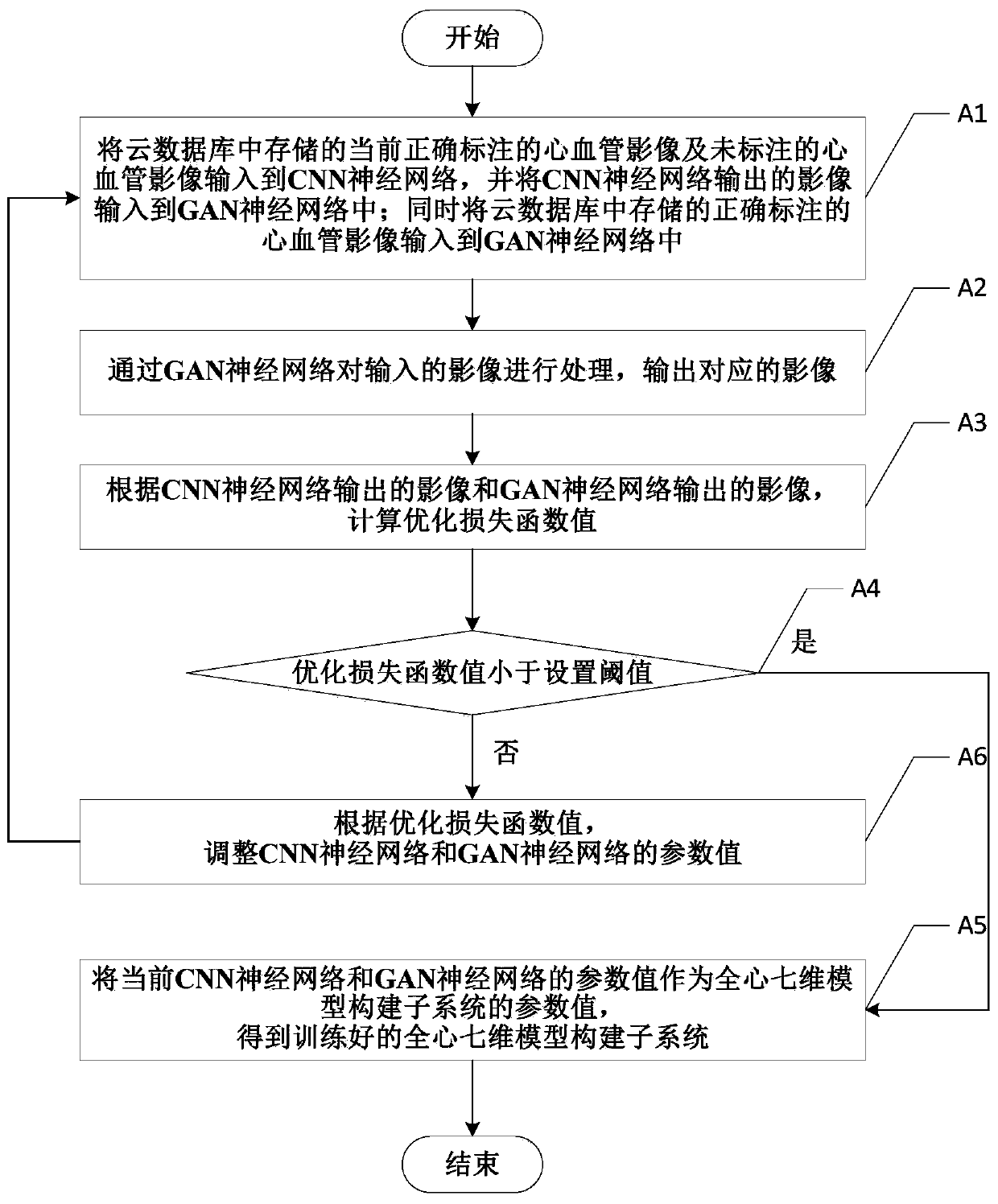
[0072] In step S1 of the embodiment of the present invention, the method for training the image data processing subsystem is as follows: input the manually correctly labeled cardiovascular images into the con...


PUM


Abstract

The invention discloses a cardiovascular image recognition system and method based on a whole-heart seven-dimensional model. The system comprises an image data processing subsystem, a cloud database and a whole-heart seven-dimensional model construction subsystem which are connected in sequence. The image data processing subsystem is used for processing an input cardiovascular image to be identified, accurately labeling the cardiovascular image and uploading the cardiovascular image to be identified to the cloud database; the cloud database is used for storing all accurately marked cardiovascular images; and the whole-heart seven-dimensional model construction subsystem is used for constructing a corresponding whole-heart seven-dimensional model according to the required cardiovascular image in the cloud database, and recognizing the cardiovascular image according to the constructed whole-heart seven-dimensional model. The cardiovascular ultrasound image can be accurately recognized, and the recognition quality is guaranteed; a high-compatibility modeling system is provided, and the method is suitable for various imaging inspection modes; the time consumed by an imaging doctor in image reading and modeling is reduced, and the burden of the imaging doctor is greatly reduced.

Description

technical field [0001] The invention belongs to the technical field of ultrasonic image recognition, and in particular relates to a cardiovascular image recognition system and method based on a seven-dimensional model of the whole heart. Background technique [0002] In the field of cardiovascular image acquisition technology, the most commonly used imaging techniques are cardiovascular ultrasound and computerized tomography (Computed Tomography, CT), and the two complement each other functionally. Among them, cardiovascular ultrasound is to use the human cardiovascular system to observe the tissue and organs, and the color ultrasound of the heart is the only inspection method that can dynamically display the structure of the heart cavity, the pulsation of the heart and hemodynamics. Commonly used methods include B-mode ultrasound, M-mode ultrasound, and Doppler ultrasound. Computed Tomography (CT) is an imaging technology that obtains cross-sectional information of an obje...

Claims


Application Information

Patent Timeline
IPC IPC(8): G16H30/20G06N3/04G06N3/08
CPCG16H30/20G06N3/08G06N3/045
Inventor 刘琦李登周翔鸿葛玲玲姚怡君
Owner SICHUAN UNIV
Why Eureka
Try Eureka