Multi-label multi-example image detection method and device, equipment and storage medium

An image detection and multi-instance technology, applied in image analysis, image data processing, capturing objects visible under the microscope, etc., can solve the problems of large number of models and cumbersome marking process, etc.

Pending Publication Date: 2020-07-28
PING AN TECH (SHENZHEN) CO LTD
View PDF0 Cites 2 Cited by
  • Summary
  • Abstract
  • Description
  • Claims
  • Application Information

AI Technical Summary

Problems solved by technology

[0004] In clinical diagnosis, the doctor will analyze the entire pathological image to obtain a diagnosis report. In most cases, it will not clearly indicate which area has which pathological characteristics in the diagnosis report. Regions need to call the corresponding model for marking, N features need N models, the number of models used is large, and the marking process is cumbersome

Method used


Image

  • Multi-label multi-example image detection method and device, equipment and storage medium
  • Multi-label multi-example image detection method and device, equipment and storage medium
  • Multi-label multi-example image detection method and device, equipment and storage medium

Examples


Embodiment Construction

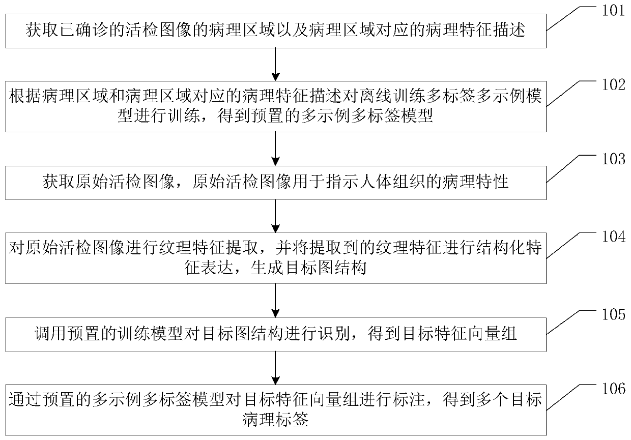
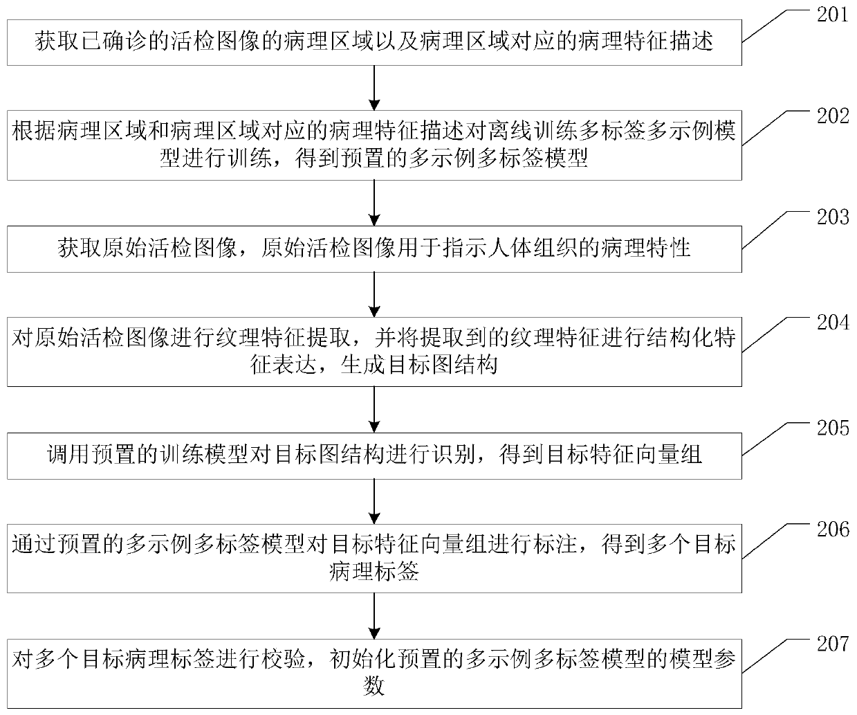
[0030] The present invention provides a multi-label and multi-instance image detection method, device, equipment and storage medium, which are used to detect pathological images in real time through a multi-label and multi-instance model, generate pathological characteristics of multiple biopsy images, and reduce the impact on pathology. The analysis time of the image is long, the analysis efficiency is improved, and the accuracy of the analysis result is improved.

[0031] In order to enable those skilled in the art to better understand the solutions of the present invention, the embodiments of the present invention will be described below with reference to the drawings in the embodiments of the present invention.

[0032] The terms "first", "second", "third", "fourth", etc. (if any) in the description and claims of the present invention and the above drawings are used to distinguish similar objects, and not necessarily Used to describe a specific sequence or sequence. It is...


PUM


Abstract

The invention relates to the field of artificial intelligence, and discloses a multi-label multi-example image detection method, device and equipment and a storage medium, which are used for reducingthe analysis duration of a pathological image and improving the accuracy of an analysis result. The method comprises the following steps: acquiring a pathological region of a diagnosed biopsy image and pathological feature description corresponding to the pathological region; training the offline training multi-label multi-example model according to the pathological region and the pathological feature description corresponding to the pathological region to obtain a preset multi-example multi-label model; acquiring an original biopsy image, wherein the original biopsy image is used for indicating pathological characteristics of human tissues; performing texture feature extraction on the original biopsy image, and performing structured feature expression on the extracted texture features togenerate a target image structure; calling a preset training model to identify the target graph structure to obtain a target feature vector group; and labeling the target feature vector group througha preset multi-example multi-label model to obtain a plurality of target pathological labels.

Description

technical field [0001] The present invention relates to the technical field of area extraction, and in particular to a multi-label and multi-instance image detection method, device, equipment and storage medium. Background technique [0002] A biopsy image is an image obtained by slicing the tissue of the diseased part of the patient and zooming in on the image under a microscope. Biopsy images directly reflect the lesions that occur inside the tissue, and are an important basis for doctors to diagnose diseases, and even the final basis for the diagnosis of some serious diseases. For example, in the diagnosis of cancer, through the observation of imaging means (X-ray, CT, MRI, etc.) diagnosis. But to make a final diagnosis, living tissue of the lesion must be extracted, observed under a microscope and biochemically tested. This process is called biopsy. [0003] Taking dermatology as an example, there are more than 3,000 common skin diseases, but only a few hundred can be...

Claims


Application Information

Patent Timeline
Patent Type & Authority Applications(China)
IPC IPC(8): G06T7/00G06K9/46G06K9/62
CPCG06V20/693G06V20/695G06V20/698G06V10/467G06V10/44G06F18/23213G06F18/24155G06F18/2411
Inventor 梁志成
Owner PING AN TECH (SHENZHEN) CO LTD
Why Eureka
Try Eureka