
Microscopic imaging device based on vertical illumination

A microscopic imaging and image acquisition device technology, applied in the field of optical imaging, can solve the problems of large image background noise and poor Z-direction resolution, etc., and achieve the effect of improving Z-direction resolution and reducing image background noise

Active Publication Date: 2020-08-25
UNIV OF SCI & TECH OF CHINA
View PDF10 Cites 5 Cited by
  • Summary
  • Abstract
  • Description
  • Claims
  • Application Information

AI Technical Summary

Problems solved by technology

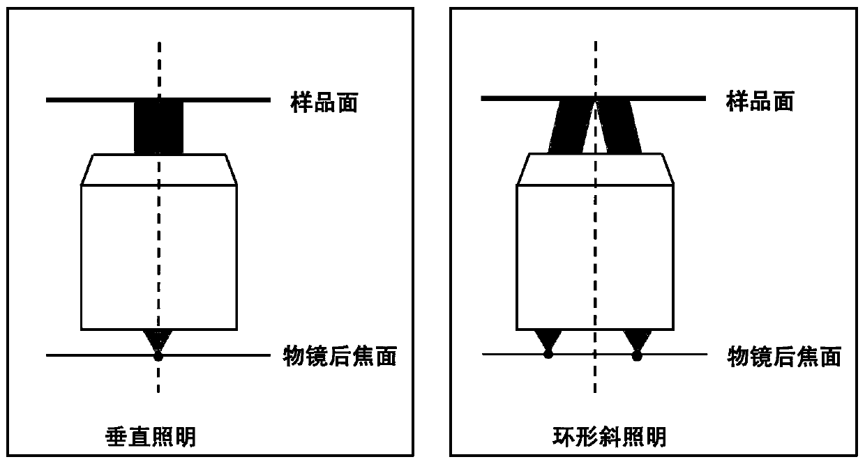
[0004] In view of this, the present invention provides a microscopic imaging device based on epi-illumination to solve the problems of poor Z-direction resolution and large image background noise in the microscopic imaging device in the prior art

Method used


Image

  • Microscopic imaging device based on vertical illumination
  • Microscopic imaging device based on vertical illumination
  • Microscopic imaging device based on vertical illumination

Examples


Embodiment Construction

[0047] As mentioned in the background technology section, when the microscopic imaging device in the prior art studies thick samples, there are problems of low Z-direction resolution and large image background noise.

[0048] The inventors found that the thick samples in the prior art have enhanced scattering and absorption of light due to their dense tissue structure, which requires higher resolution in the Z direction of the imaging technology; at the same time, the unscattered light in the sample and multiple Scattered light will lead to a bright background, how to reduce the background and improve the contrast of the image becomes the key.

[0049] The current main tools for imaging thick samples in the state of the art include: confocal microscopy, two-photon fluorescence microscopy, and light sheet microscopy. These three microscopic imaging techniques are all based on fluorescent labels to generate image contrast. Fluorescent labels will affect the structure of biologic...


PUM

PropertyMeasurementUnit
wavelengthaaaaaaaaaa
widthaaaaaaaaaa

Abstract

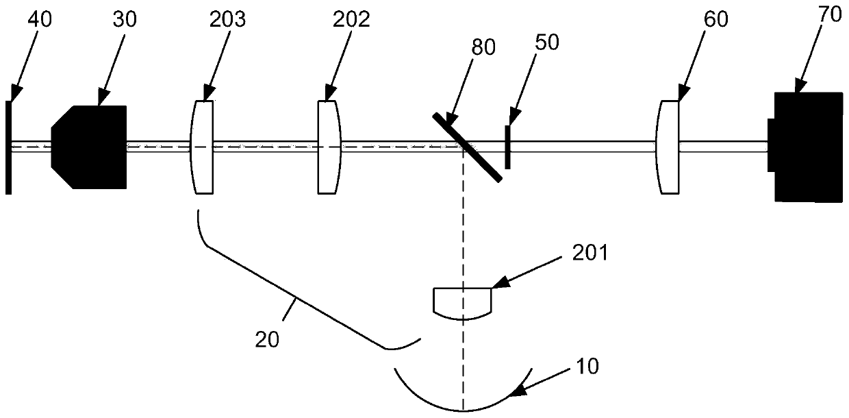
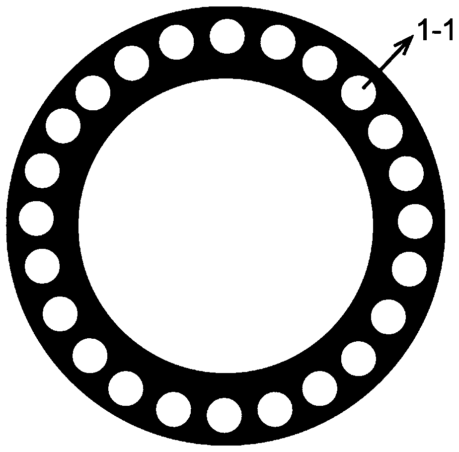
The invention provides a microscopic imaging device based on incident illumination. The microscopic imaging device comprises an annular light source, a first focusing module, a microscope objective, asample table, an annular attenuation sheet, a second focusing module and a first image collection device, microscopic imaging device, according to the invention, the back scattering signals of the thick sample are acquired, the interference of the system background and the diffuse reflection of the sample on the weak back scattering signals is reduced through the annular attenuation sheet, and the clear back scattering signals are acquired, so that the Z-direction resolution of the thick sample of the microscopic imaging device is improved, and the background noise of the image is reduced.

Description

technical field [0001] The invention relates to the technical field of optical imaging, in particular to a microscopic imaging device based on epi-illumination. Background technique [0002] The three-dimensional microscopic imaging technology with high spatio-temporal resolution and high contrast is of great significance to the study of thick samples such as model organisms. Realizing the visualization of various tissues in model organisms with cell-level resolution is very useful in genetic biology and neurobiology. prospect. [0003] However, when the microscopic imaging technology in the prior art studies thick samples, the resolution in the Z direction is low and the image background noise is large. Contents of the invention [0004] In view of this, the present invention provides a microscopic imaging device based on epi-illumination to solve the problems of poor Z-direction resolution and large image background noise in the microscopic imaging device in the prior a...

Claims


Application Information

Patent Timeline
Patent Type & Authority Applications(China)
IPC IPC(8): G02B21/00G02B21/06G02B21/24G02B21/36G01N21/64G01N21/84
CPCG01N21/6458G01N21/84G02B21/0032G02B21/06G02B21/241G02B21/361
Inventor 储开芹扎克时瑞洁
Owner UNIV OF SCI & TECH OF CHINA
Learn More