
Fluorescence control method for molecular labeling of biological tissues with organic fluorescent dyes

A technology of fluorescent dye and control method, which is applied in the field of fluorescence control of organic fluorescent dye molecules to label biological tissues, can solve the problems of reducing the density of conjugated π electrons of organic fluorescent molecules, difficulty of accurate three-dimensional registration, and interference of imaging background fluorescence, etc. The effect of suppressing the interference of background fluorescence, reversible quenching, and convenient reactivation

Active Publication Date: 2019-05-31
HUAZHONG UNIV OF SCI & TECH
View PDF2 Cites 0 Cited by
  • Summary
  • Abstract
  • Description
  • Claims
  • Application Information

AI Technical Summary

Problems solved by technology

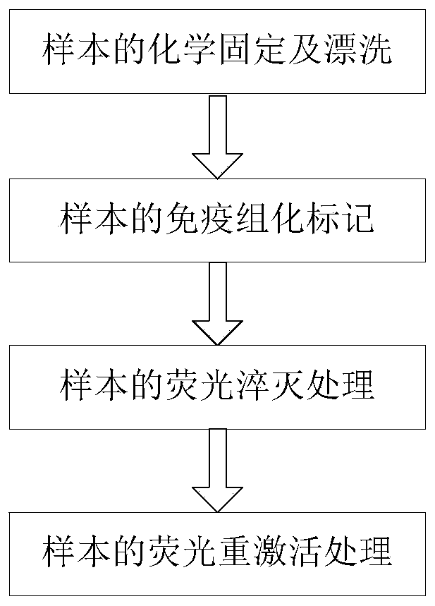
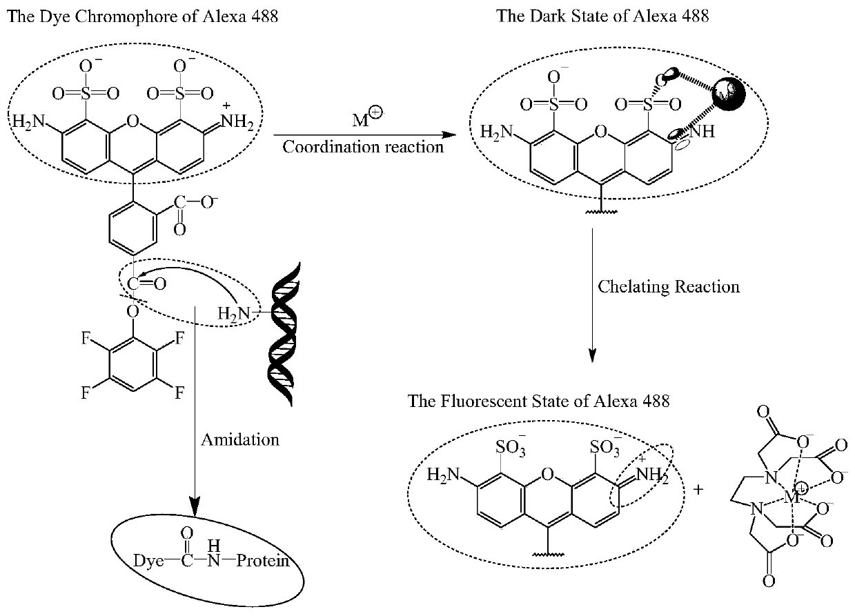
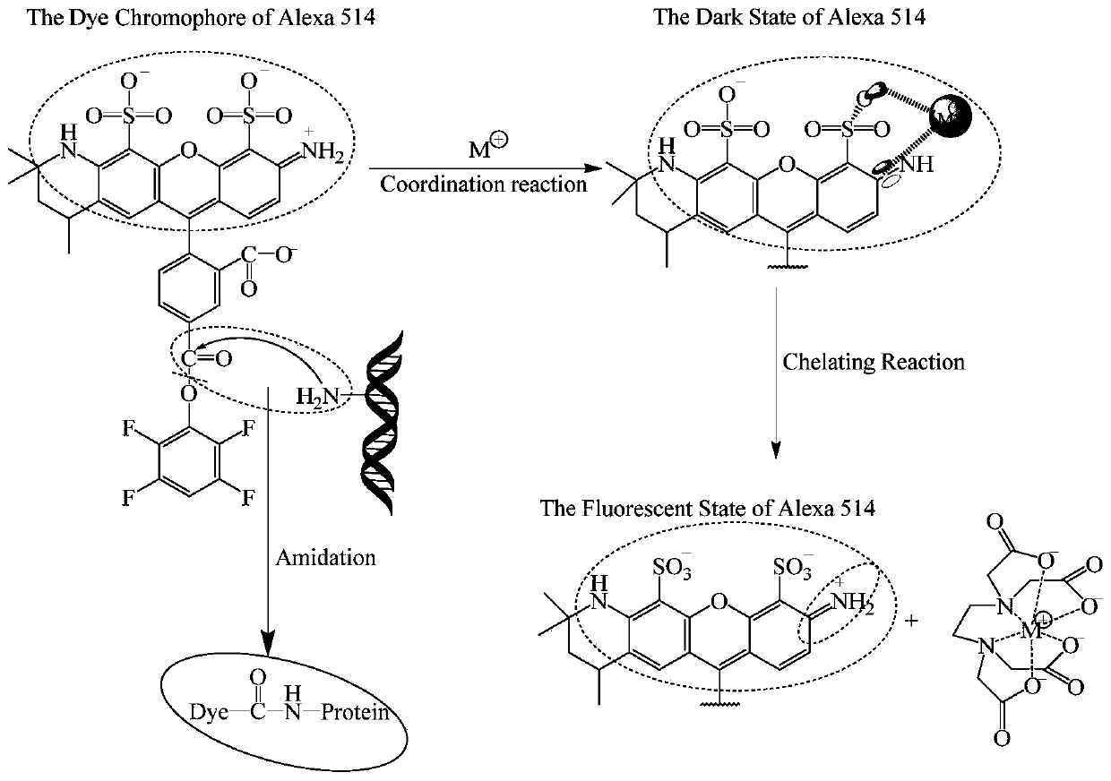
[0005] In view of the above defects or improvement needs of the prior art, the present invention provides a method for controlling the fluorescence of organic fluorescent dye molecularly labeled biological tissues, the purpose of which is to reduce organic fluorescence by combining metal ions with organic fluorescent dye molecules to form a six-membered ring The conjugated π electron density of the molecule can reversibly quench the fluorescence of the organic fluorescent dye molecule; then use the metal ion chelating agent to form a coordination bond with the metal ion, destroy the combination of the metal ion and the organic fluorescent dye molecule, and restore its conjugated π Electronic structure, thereby reactivating the fluorescence of organic fluorescent dye molecules, through the fluorescence control method of the present invention, and applying it to tomographic imaging technology, thus solving the problem of incomplete quenching in the prior art physical tomographic technology that leads to imaging Technical problems such as background fluorescence interference, low image acquisition throughput, low resolution in the Z direction, and difficulty in accurate 3D registration

Method used


Image

  • Fluorescence control method for molecular labeling of biological tissues with organic fluorescent dyes
  • Fluorescence control method for molecular labeling of biological tissues with organic fluorescent dyes
  • Fluorescence control method for molecular labeling of biological tissues with organic fluorescent dyes

Examples


Embodiment 1

[0053] (1) Fix the perfused mouse brain with 4% PFA solution and rinse with PBS solution;

[0054] (2) Immunohistochemically label the fixed and rinsed 100-micron mouse brain tissue slices with Alexa 488 and rinse with PBS solution: first, fix the 100-micron slices with 20% DMSO and 0.2% Triton X-100 in PBS solution Punch holes with the rinsed mouse brain tissue slices for 12 hours, then add 10% serum to block for 12 hours, rinse with PBS for 1 hour / 3 times, then add the primary antibody at a ratio of 500:1 at 37°C in the dark Incubate with shaking for 2 days, then incubate with shaking at 37°C for 2 days in the dark, then add the secondary antibody with Alexa 488 at a ratio of 800:1, incubate with shaking for 8 hours at 4°C in the dark, and rinse with PBS for 1 hour / 3 times to obtain Alexa 488 immunohistochemically labeled 100 micron mouse brain tissue slice;

[0055] (3) Soak 100 micron mouse brain tissue slices immunohistochemically labeled with Alexa 488 in 10mmol / L FeSO ...

Embodiment 2

[0059] (1) Fix the perfused mouse brain with 4% PFA solution and rinse with PBS solution;

[0060] (2) Immunohistochemically label the fixed and rinsed 100-micron mouse brain tissue slices with Alexa 488 and rinse with PBS solution: first, fix the 100-micron slices with 20% DMSO and 0.2% Triton X-100 in PBS solution Punch holes with the rinsed mouse brain tissue slices for 12 hours, then add 10% serum to block for 12 hours, rinse with PBS for 1 hour / 3 times, then add the primary antibody at a ratio of 500:1 at 37°C in the dark Incubate with shaking for 2 days, then incubate with shaking at 37°C for 2 days in the dark, then add the secondary antibody with Alexa 488 at a ratio of 800:1, incubate with shaking for 8 hours at 4°C in the dark, and rinse with PBS for 1 hour / 3 times to obtain Alexa 488 immunohistochemically labeled 100 micron mouse brain tissue slice;

[0061] (3) Soak 100 micron mouse brain tissue slices immunohistochemically labeled with Alexa 488 in 100mmol / L FeCl...

Embodiment 3

[0065] (1) The perfused 100-micron mouse brain tissue was fixed with 4% PFA solution and rinsed with PBS solution;

[0066](2) Immunohistochemically label the fixed and rinsed 100-micron mouse brain tissue slices with Alexa 514 and rinse with PBS solution: first, fix the 100-micron slices with 20% DMSO and 0.2% Triton X-100 in PBS solution Punch holes with the rinsed mouse brain tissue slices for 12 hours, then add 10% serum to block for 12 hours, rinse with PBS for 1 hour / 3 times, then add the primary antibody at a ratio of 500:1 at 37°C in the dark Incubate with shaking for 2 days, then incubate with shaking at 37°C for 2 days in the dark, then add the secondary antibody with Alexa 514 at a ratio of 800:1, incubate with shaking at 4°C for 8 hours in the dark, and rinse with PBS for 1 hour / 3 times to obtain Alexa 514 immunohistochemically labeled 100 micron mouse brain tissue slice;

[0067] (3) Use 200mmol / L Fe 2 (SO 4 ) 3 Imaging was performed after the solution was que...


PUM


Abstract

The invention discloses a fluorescence control method used in labeling biological tissues with organic fluorescent dye molecules. According to the fluorescence control method, hexatomic rings are formed via combination of metal ions with the organic fluorescent dye molecules, and quenching of the fluorescence of the organic fluorescent dye molecules is realized; coordination bonds are formed by a metal ion chelating agent with metal ions, combination of the metal ions with the organic fluorescent dye molecules is destroyed, and reactivation of the fluorescence of the organic fluorescent dye molecules is realized. The fluorescence control method is capable of realizing complete fluorescence quenching, and convenient and controllable reactivation. The fluorescence control method used in labeling biological tissues with organic fluorescent dye molecules is combined with a wide-field imaging system, so that chemical chromatography imaging of samples labeled with the fluorescent dye molecules is realized, and rapid chemical chromatography imaging of large-size biological tissues labeled with organic fluorescent molecules is realized.

Description

technical field [0001] The invention belongs to the field of biological imaging, and more specifically relates to a fluorescence control method for molecularly marking biological tissues with organic fluorescent dyes. Background technique [0002] Obtaining the detailed spatial distribution of molecules and proteins in biological tissues is an important basis for understanding the relationship between the structure and function of organisms. Fluorescent probe labeling technology can label specific molecules and proteins. In order to better study the spatial distribution and function of these molecules and proteins in biological tissues, a variety of fluorescent probe labeling methods in the same organism have been developed. It is widely used in the fields of biology and medicine. [0003] For optical imaging of biological tissues labeled with fluorescent probes, there are mainly two existing physical tomography techniques: one is optical tomography, which optically suppres...

Claims


Application Information

Patent Timeline
Patent Type & Authority Patents(China)
IPC IPC(8): G01N21/64G01N1/30
CPCG01N1/30G01N21/6428G01N2001/302G01N2021/6432G01N2021/6439
Inventor 骆清铭周宏福刘秀丽刚亚栋曾绍群
Owner HUAZHONG UNIV OF SCI & TECH
Learn More