Ki67 cell nucleus counting method and system based on pathological immunohistochemistry

A technique of immunohistochemistry and counting method, applied in the field of Ki67 cell nucleus counting method and system, can solve the problems of large workload, waste of medical resources, time-consuming and laborious, etc., and achieve the effect of accurate diagnosis results

Pending Publication Date: 2020-08-25
QIANFOSHAN HOSPITAL OF SHANDONG +1
View PDF7 Cites 9 Cited by
  • Summary
  • Abstract
  • Description
  • Claims
  • Application Information

AI Technical Summary

Problems solved by technology

On the one hand, the interpretation results are completely affected by subjectivity and experience, and the interpretation of the same slice varies greatly by different people; on the other hand, there is a shortage of pathologists, and the relative workload is relatively large. waste of medical resources

Method used


Image

  • Ki67 cell nucleus counting method and system based on pathological immunohistochemistry
  • Ki67 cell nucleus counting method and system based on pathological immunohistochemistry
  • Ki67 cell nucleus counting method and system based on pathological immunohistochemistry

Examples


Embodiment 1

[0046] Embodiment 1, this embodiment provides a method for counting Ki67 cell nuclei based on pathological immunohistochemistry;

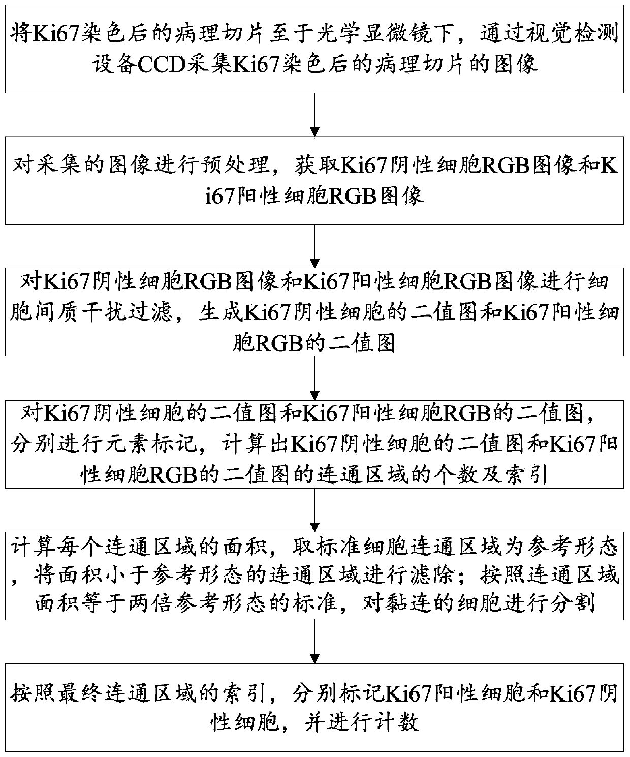
[0047] Such as figure 1 As shown, the method of counting Ki67 cell nuclei based on pathological immunohistochemistry, including:

[0048] S101: image acquisition step: place the Ki67-stained pathological section under an optical microscope, and collect the image of the Ki67-stained pathological section through a visual detection device CCD;

[0049] S102: Image preprocessing step: preprocessing the collected images to obtain RGB images of Ki67-negative cells and RGB images of Ki67-positive cells;

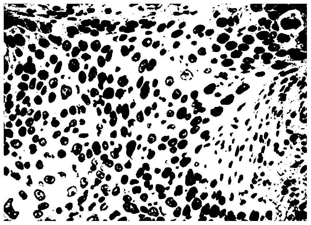
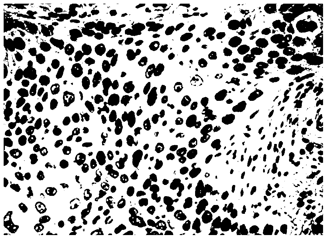
[0050] S103: Intercellular interference processing step: performing intercellular interference filtering on the RGB images of Ki67-negative cells and RGB images of Ki67-positive cells to generate a binary image of Ki67-negative cells and a binary image of RGB of Ki67-positive cells;

[0051] S104: Mark the binary image of Ki67-negative cells and the bina...

Embodiment 2

[0096] Embodiment 2, this embodiment provides a Ki67 cell nucleus counting system based on pathological immunohistochemistry;

[0097] Ki67 cell nucleus counting system based on pathological immunohistochemistry, including:

[0098] An image acquisition module configured to: place the Ki67-stained pathological section under an optical microscope, and collect an image of the Ki67-stained pathological section through a visual detection device CCD;

[0099] An image preprocessing module, which is configured to: preprocess the collected images, and obtain RGB images of Ki67-negative cells and RGB images of Ki67-positive cells;

[0100] The intercellular interference processing module is configured to: perform intercellular interference filtering on the Ki67 negative cell RGB image and the Ki67 positive cell RGB image, and generate a binary image of the Ki67 negative cell and a binary image of the Ki67 positive cell RGB;

[0101] The marking module is configured to: perform elemen...

Embodiment 3

[0104] Embodiment 3. This embodiment also provides an electronic device, including a memory, a processor, and computer instructions stored in the memory and run on the processor. When the computer instructions are executed by the processor, the computer instructions in Embodiment 1 are completed. described method.


PUM


Abstract

The invention discloses a Ki67 cell nucleus counting method and system based on pathological immunohistochemistry, and the method comprises the steps: placing a pathological section after Ki67 staining under an optical microscope, and collecting an image of the pathological section after Ki67 staining through visual detection equipment CCD; obtaining a Ki67 negative cell RGB image and a Ki67 positive cell RGB image; carrying out intercellular substance interference filtering on the RGB image to generate a binary image of Ki67 negative cells and a binary image of Ki67 positive cell RGB; carrying out element marking on the binary image, and calculating the number and indexes of connected regions of the binary image; calculating the area of each connected region, taking a standard cell connected region as a reference form, and filtering the connected region with the area smaller than the reference form; segmenting the adherent cells according to the standard that the area of the connectedregion is equal to twice of the reference form; marking Ki67 positive cells and Ki67 negative cells according to the index of the final connected region, and performing counting.

Description

technical field [0001] The present disclosure relates to the field of medical technology, in particular to a method and system for counting Ki67 cell nuclei based on pathological immunohistochemistry. Background technique [0002] The statements in this section merely mention background art related to the present disclosure and do not necessarily constitute prior art. [0003] Ki67 is a nuclear antigen (non-histone) that exists in each cycle of cell division. In pathology, immunohistochemical methods are usually used to mark its expression in the nucleus, so as to understand at a certain point in time. How many cells are in the stage of division and proliferation, and how many cells are in the quiescent stage, are a very meaningful indicator for judging the activity of tumor cells. Clinically, it is most commonly used in the histopathological diagnosis of breast cancer, neuroendocrine tumors, meningiomas, etc. It is of great value in judging benign and malignant tumors, rec...

Claims


Application Information

Patent Timeline
Patent Type & Authority Applications(China)
IPC IPC(8): G06T7/00G06T7/136G06T7/187G06T7/62G01N15/14
CPCG06T7/0012G06T7/187G06T7/62G06T7/136G01N15/1429G06T2207/10061G06T2207/30024G06T2207/30242G01N2015/1486
Inventor 孙青张国霞成玉霞张欣欣胡维荣蔡东兴
Owner QIANFOSHAN HOSPITAL OF SHANDONG
Why Eureka
Try Eureka