Supercharge Your Innovation With Domain-Expert AI Agents!

A Multi-contrast Image Extraction Method for X-ray Diffraction Enhanced Imaging

A technique for enhancing imaging, extraction methods, applied in the field of X-ray imaging

Active Publication Date: 2022-03-29
HEFEI UNIV OF TECH
View PDF12 Cites 0 Cited by
  • Summary
  • Abstract
  • Description
  • Claims
  • Application Information

AI Technical Summary

Problems solved by technology

This is one of the limitations of the current multi-map statistical method

Method used


Image

  • A Multi-contrast Image Extraction Method for X-ray Diffraction Enhanced Imaging
  • A Multi-contrast Image Extraction Method for X-ray Diffraction Enhanced Imaging
  • A Multi-contrast Image Extraction Method for X-ray Diffraction Enhanced Imaging

Examples


Embodiment Construction

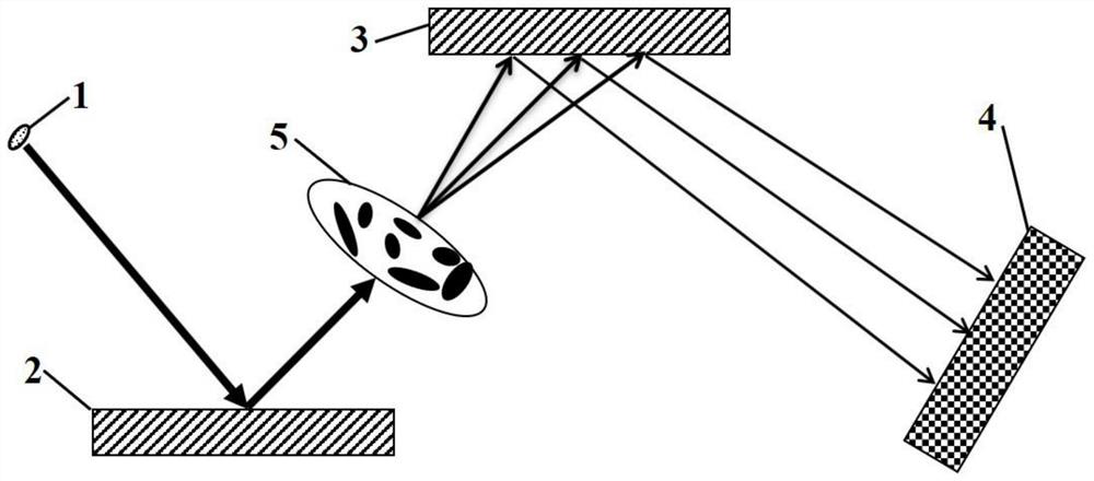
[0043] In this example, see figure 1 , setting an X-ray diffraction enhanced imaging device composed of X-ray source 1, monochromatic crystal 2, analysis crystal 3 and detector 4; as figure 1 As shown, the X-ray propagation direction is the Z axis; the X-ray source 1, the monochromatic crystal 2, the imaged object 5, the analysis crystal 3 and the detector 4 are sequentially arranged along the Z axis; the X-ray diffraction enhanced imaging The multi-contrast information extraction method of is carried out as follows:

[0044] Step 1. Set the relative position of each device, satisfying: 01 2 3 , where Z 1 is the relative distance between the X-ray source 1 and the monochromatic crystal 2 along the Z axis; Z 2 is the relative distance between the X-ray source 1 and the analysis crystal 3 along the Z axis; Z 3 is the relative distance between the X-ray source 1 and the detector 4 along the Z-axis;

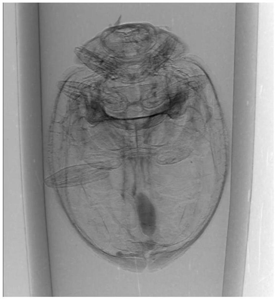
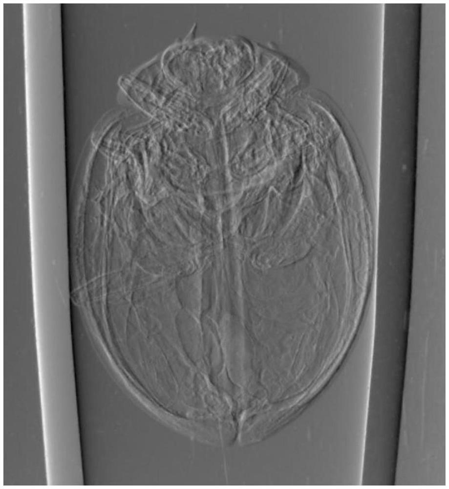
[0045] Step 2. Obtain the background projection image:

[0046] Step 2.1, t...


PUM


Abstract

The invention discloses a multi-contrast image extraction method for X-ray diffraction enhanced imaging, which is applied to diffraction enhanced imaging formed by sequentially arranging an X-ray source, a monochromatic crystal, an analysis crystal and a detector along the X-ray propagation direction. In the device; X-rays are diffracted after incident on a monochromatic crystal, and the outgoing monochromatic collimated X-rays penetrate the imaged object and then enter the analysis crystal and are diffracted. After the outgoing X-rays are incident on the detector, the intensity of the X-rays It is detected and recorded by the detector; taking the normal of the diffraction surface of the analysis crystal as the rotation axis, the projection images of the different angular positions of the analysis crystal along the rotation axis are recorded by the detector, and the detector is processed by the proposed multi-contrast image extraction method The recorded projection image can obtain the absorption image, refraction image, scattering image, skewness image and kurtosis image of the imaged object. The invention can solve the problem of accurate and quantitative extraction of the skewness image and the kurtosis image of the imaged object.

Description

technical field [0001] The invention relates to the field of X-ray imaging methods, in particular to a multi-contrast image extraction method for X-ray diffraction enhanced imaging. Background technique [0002] After more than 100 years of continuous development and improvement, X-ray imaging technology has been widely used in many fields such as public security inspection, clinical medical diagnosis and treatment, nondestructive testing, and material science. As the mainstream of the current X-ray imaging technology, the X-ray absorption contrast imaging method can obtain good image quality when it is applied to objects mainly composed of high atomic number elements. However, for objects mainly composed of low atomic number elements, including organic composite materials, human soft tissues, etc., their intensity attenuation to X-rays is very weak. Using the absorption contrast imaging technology, the image quality obtained is very poor, and the accurate identification of...

Claims


Application Information

Patent Timeline
Patent Type & Authority Patents(China)
IPC IPC(8): G01N23/207
CPCG01N23/207
Inventor 王志立陈恒
Owner HEFEI UNIV OF TECH
Features
  • R&D
  • Intellectual Property
  • Life Sciences
  • Materials
  • Tech Scout
  • Unparalleled Data Quality
  • Higher Quality Content
  • 60% Fewer Hallucinations
Learn More