
Bimodal microimaging system and method

A technology for microscopic imaging and imaging subsystems, applied in microscopes, fluorescence/phosphorescence, instruments, etc.

Active Publication Date: 2020-09-01
PEKING UNIV
View PDF17 Cites 14 Cited by
  • Summary
  • Abstract
  • Description
  • Claims
  • Application Information

AI Technical Summary

Problems solved by technology

In traditional optical diffraction tomography, only lipid droplets, chromosomes and mitochondria have been identified and calibrated in combination with wide-field fluorescence imaging.

Method used


Image

  • Bimodal microimaging system and method
  • Bimodal microimaging system and method
  • Bimodal microimaging system and method

Examples


Embodiment Construction

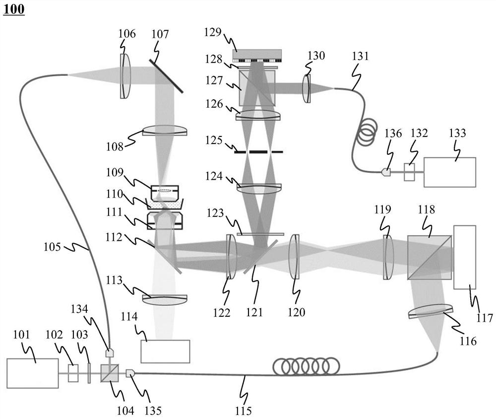
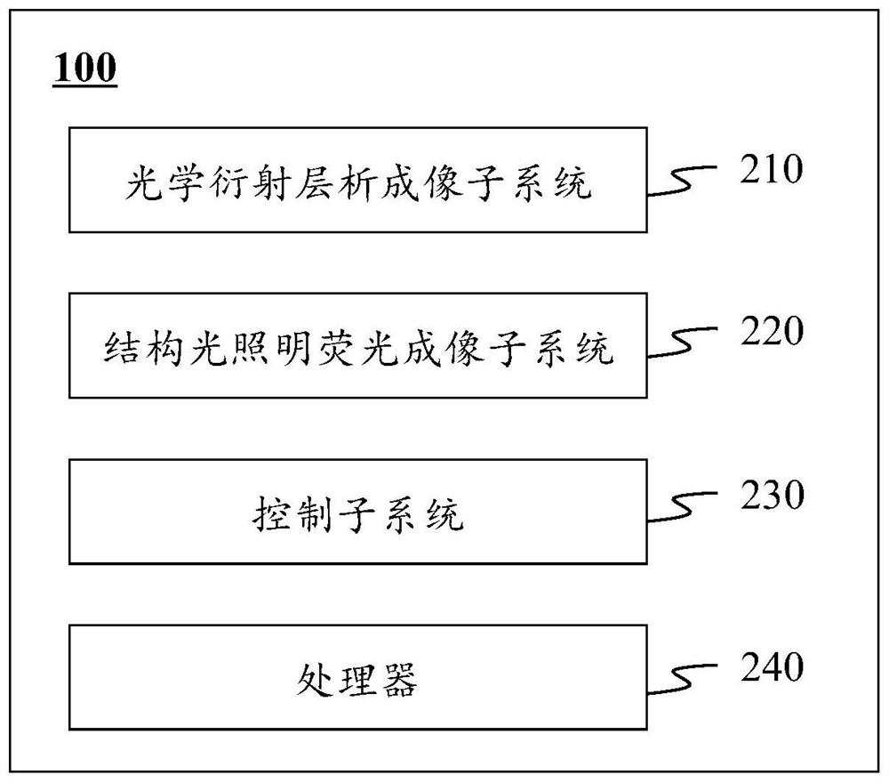
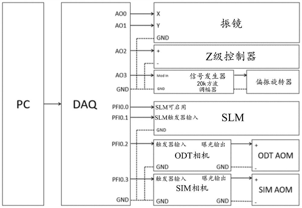
[0050] In order to more clearly describe the technical solutions of the embodiments of the present application, the following will briefly introduce the drawings that need to be used in the description of the embodiments. Obviously, the drawings in the following description are just some examples or embodiments of the application. For those of ordinary skill in the art, without creative work, the application can be applied to the application according to these drawings. Other similar scenarios. Unless it is obvious from the language environment or otherwise stated, the same reference numerals in the figures represent the same structure or operation.

[0051] It should be understood that the "system", "device", "unit" and / or "module" used herein are a method for distinguishing different components, elements, parts, parts or assemblies of different levels. However, if other words can achieve the same purpose, they can be replaced by other expressions.

[0052] As shown in the prese...


PUM

PropertyMeasurementUnit
Horizontal resolutionaaaaaaaaaa
Vertical resolutionaaaaaaaaaa
Horizontal resolutionaaaaaaaaaa

Abstract

The embodiment of the invention discloses a bimodal microimaging system and method; the bimodal microimaging system comprises an optical diffraction tomography subsystem and a structured light illumination fluorescence imaging subsystem; the optical diffraction tomography subsystem is used for carrying out unmarked optical diffraction tomography based on the first laser so as to obtain an opticaldiffraction tomography image of the sample; the structured light illumination fluorescence imaging subsystem is used for performing fluorescence imaging based on the second laser so as to obtain a structured light illumination fluorescence image of the sample; the bimodal microimaging system comprises a first light source and a second light source which are independent of each other, wherein the first light source is used for emitting the first laser, and the second light source is used for emitting the second laser.

Description

Technical field [0001] This application relates to the field of microscopic imaging technology, and in particular to a dual-modality microscopic imaging system and method. Background technique [0002] Since the 21st century, the vigorous development of biology and medicine has continuously improved mankind’s understanding of the nature of life and greatly promoted the development of human civilization. With the continuous development of biomedical research, its cutting-edge research has penetrated into the cell, trying to decrypt the life process on the molecular scale. Optical microscopy is a powerful tool for modern molecular biology research. Its development promotes the continuous progress of human observation and understanding of life phenomena. An optical microscope is a device that uses the interaction of light and matter to present the microstructure of an object. Since the invention of the optical microscope, it has been the most commonly used tool in the biomedical f...

Claims


Application Information

Patent Timeline
IPC IPC(8): G02B21/00G02B21/06G02B21/36G01N21/64
CPCG02B21/0048G02B21/0076G02B21/06G02B21/361G01N21/6458
Inventor 施可彬陈良怡董大山黄小帅李柳菊毛珩王爱民
Owner PEKING UNIV
Features
  • Generate Ideas
  • Intellectual Property
  • Life Sciences
  • Materials
  • Tech Scout
  • Unparalleled Data Quality
  • Higher Quality Content
  • 60% Fewer Hallucinations
Learn More