[0015]It is an object of the present invention to provide in vivo endoscopic methods for implementation of AF
microscopy, without the use of exogenous agents, to effectively (with resolution sufficient to image nuclei) visualize and categorize various abnormal
esophageal tissue forms.
[0016]Another object is to provide apparatuses capable of in vivo endoscopic methods for implementation of AF (without exogenous agents)
microscopy to effectively visualize and categorize various abnormal
esophageal tissue forms.
[0019]A microscope
system described in US20070160279(A1), incorporated herein by reference, has been used to determine the most effective excitation-emission combinations for imaging changes of structural and biochemical characteristics in esophagus tissue [1]. Ultra violet (UV) wavelengths were implemented in this platform as the excitation sources for
ex vivo human esophagus tissue biopsies. The present inventors have made the discovery that imaging of the superficial tissue
microstructure can be achieved using conventional
wide field autofluorescence microscopy under excitation with UV light of less than 400 nm. Although the exact mechanism is still not completely clear, they hypothesize that this becomes possible via two main mechanisms. The first mechanism now discussed is that the excitation UV light only penetrates tissue to about 100 μm or less and as a result, a sufficient amount of the
fluorescence signal produced in the superficial tissue layer can be contained within the thickness of the
image plane of the microscope, thus the out of focus signal (background) is sufficiently reduced to allow the formation of
high contrast images of tissue
microstructure using the native fluorescence (
autofluorescence) of the tissue cells and
intracellular components. There is no need for a pinhole because substantially all of the AF is produced within this superficial tissue layer that has thickness comparable to the thickness of the
image plane. This
hypothesis is supported by additional experimental results that the contrast of AF images obtained with excitation wavelengths longer that about 300 nm is lower to that when the
excitation wavelength in shorter than about 300 nm, suggesting that the
photon penetration depth for shorter UV
wavelength is also shorter giving rise to a higher
image contrast. The second mechanism now discussed is that there is sufficient variability in the concentration of chromophores (that can be photo-excited with UV light) contained within subcellular and
intracellular components so that the microscopic autofluorescence image of the tissue reveals the
microstructure and organization of the superficial layer in a similar way to that provided by H&E
staining.
[0020]Further, since the present invention provides much more light for imaging than the
confocal microscope, it is able to produce an autofluorescence image in much less time (˜10 microseconds or less). This reduced time required to collect enough light to produce an adequate image enables use of autofluorescence microscopy in vivo. The limit on the duration of the
image acquisition window is that the image must be collected in an
exposure time in which the
target tissue (such as esophagus) and / or instrument (such as micro-
endoscope) moves no more that the resolution of the optical
system. Thus, if the desired resolution of the optical system is 1
micrometer, then the time it takes the
target tissue to move 1
micrometer is the limit of the duration of the
image acquisition window. The resolution S is equal to the speed, in not all, that the tissue is moving times the limit on the
exposure time. Thus, if S=1
micrometer and the combined speed of motion is on the order of V=5
millimeter / second, then the
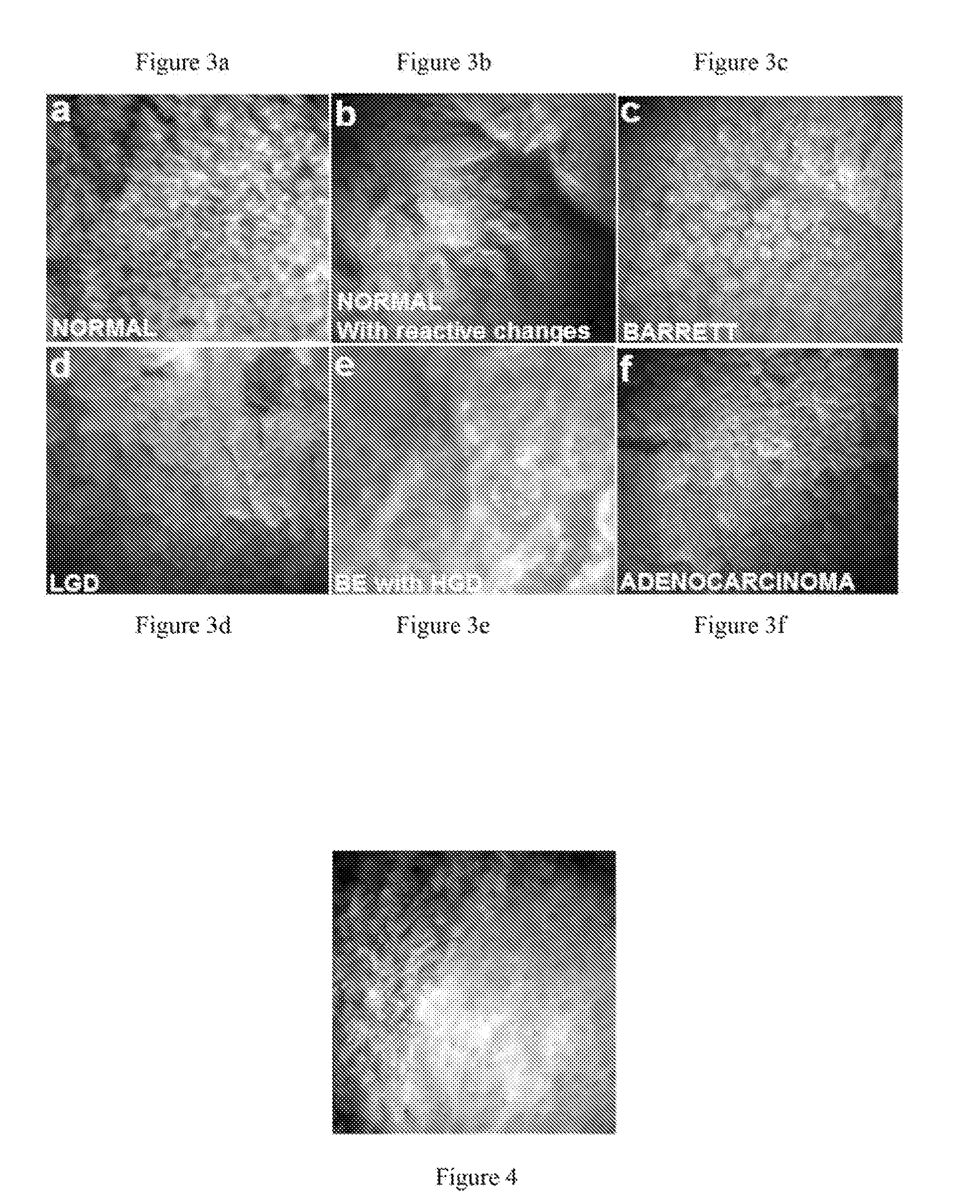
time duration limit is 200 μsec. This technology does not require exogenous agents and is non-destructive to the sample. The present invention provides a minimally (or non) invasive method to visualize and categorize various abnormal
esophageal tissue forms such as Barrett's esophagus (BE), low-grade
dysplasia (LGD), high-grade
dysplasia (HGD), and
adenocarcinoma (ADC) in vivo and in real time. Endoscopic implementations are provided.
[0021]The invention herein utilizes the AF optical signal and provides resolution sufficient to image nuclei. This technology thus bypasses the complications of contrast agents while still obtaining
diagnostic information sufficient to meet the current Hematoxylin and
Eosin (HE)
Staining Protocol, which is sometimes referred to in the field as the H&E
gold standard. Embodiments of the present invention have been successfully implemented into endoscopes. A prototype
zoom video
endoscope probe was originally designed to operate in
contact mode and is equipped with a
white light guide, objective lens, and CCD encased at the tip of the endoscope probe. The probe distal end outer
diameter is 3.2 mm and has a 300×300 μm
field of view. The design has previously been shown to image
cell nuclei with
methylene blue
contrast enhancement [34-36]. The present invention is able to acquire and categorize AF images of tissue without the use of contrast agents or hardware modification.

