Blood vessel lumen automatic segmentation method based on deep learning

A technology of deep learning and automatic segmentation, which is applied in the field of image processing, can solve problems such as inability to segment images, and achieve the effects of high accuracy, high resolution, and abstract feature extraction capabilities

Pending Publication Date: 2020-09-04
SUZHOU PULSE RONGYING MEDICAL TECH CO LTD
View PDF10 Cites 9 Cited by
  • Summary
  • Abstract
  • Description
  • Claims
  • Application Information

AI Technical Summary

Problems solved by technology

[0005] In view of the above problems, the present invention provides an automatic segmentation method of blood vessel lumen based on deep learning, which can solve the problem that the image cannot be segmented due to speckle noise, image artifacts and partial vascular wall calcification shadows, and can quickly obtain accurate Segmentation results with high accuracy and high degree of automation

Method used


Image

  • Blood vessel lumen automatic segmentation method based on deep learning
  • Blood vessel lumen automatic segmentation method based on deep learning
  • Blood vessel lumen automatic segmentation method based on deep learning

Examples


Embodiment Construction

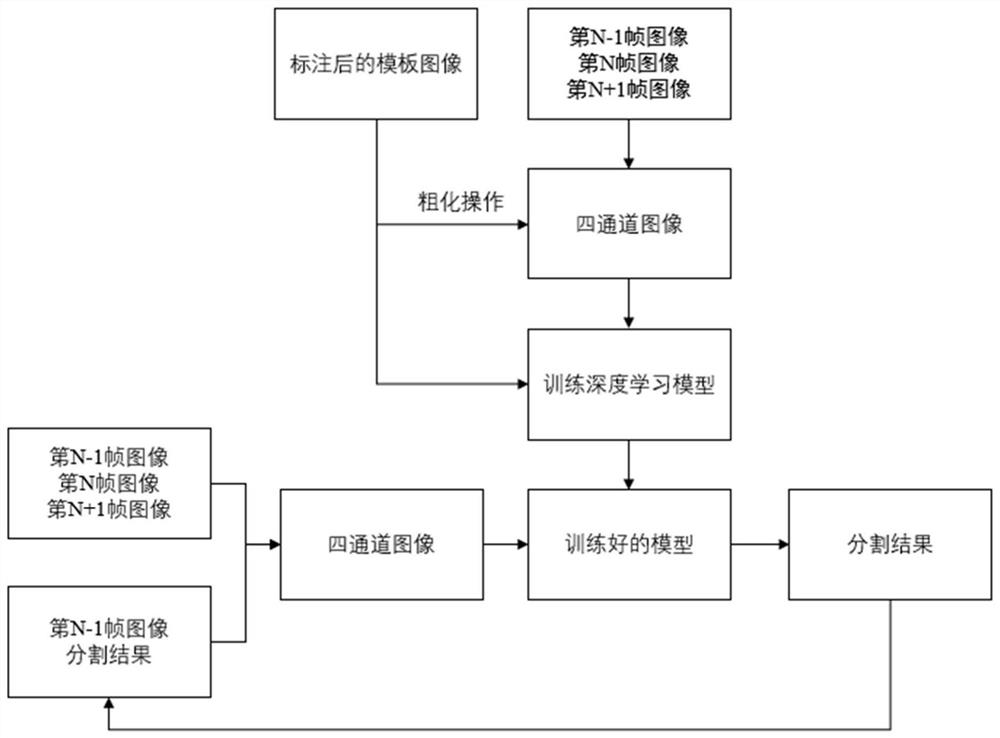
[0024] Such as Figure 1 to Figure 5 As shown, a deep learning-based automatic segmentation method for blood vessel lumen, which includes the following steps:

[0025] S1. Obtain the complete image sequence of IVUS;

[0026] S2. Manually draw the lumen-intima interface and the media-adventitia interface. Since there may be hundreds of cases and thousands of frames of images during training, the original images with representative frames are selected from the complete IVUS images. Annotate, obtain the original image and its annotated template image, and establish a training set and a test sample set; wherein, the template image contains images of speckle noise, vascular branches, image artifacts, and partial vascular wall calcification shadows;

[0027] S3. Model training stage: Affine transformation and non-rigid transformation are performed on the template image marked in the training set to obtain a roughened template image, and then the original image of the current frame ...


PUM


Abstract

The invention provides a blood vessel lumen automatic segmentation method based on deep learning. An accurate segmentation result can be rapidly obtained in real time. The method comprises the following steps: S1, acquiring an IVUS image; s2, after an original image and the marked template image are obtained, establishing a training set and a test sample set; s3, coarsening the template image marked by the training set to obtain a template image, wherein the current frame, the previous frame and the next frame of the training set and the coarsened template image form a four-channel image; s4,enabling the established deep learning segmentation model to adopt a network structure with residual connection, and inputting the template image marked in the S2 and the four-channel image constructed in the S3 into the model to obtain a trained network; and S5, forming a four-channel image by the previous frame segmentation result and the current frame, the previous frame and the next frame of images of the test sample set, inputting the four-channel image into a network for segmentation, and finally obtaining a lumen-intima interface and a middle-outer membrane interface segmented by the current frame.

Description

technical field [0001] The invention relates to the technical field of image processing, in particular to an automatic segmentation method of blood vessel lumen based on deep learning. Background technique [0002] Intravascular imaging Intravascular ultrasound (IVUS) uses high-frequency ultrasound to present high-resolution images in blood vessels, which can comprehensively analyze the conditions of blood vessel walls and lumens. It is currently the most commonly used intravascular imaging technology in clinical practice. , which can perform high-precision lumen segmentation on intravascular ultrasound images, determine the lumen-intima interface and the media-adventitia interface, and can obtain detailed lumen and plaque information to help doctors make clinical diagnoses. At the same time, Based on the segmentation results, a high-precision three-dimensional model of blood vessels can be established to help improve the accuracy of the virtual blood flow fraction calculati...

Claims


Application Information

Patent Timeline
Patent Type & Authority Applications(China)
IPC IPC(8): G06T7/00G06T7/10
CPCG06T7/0012G06T7/10G06T2207/10132G06T2207/30101G06T2207/20081G06T2207/20084
Inventor 李莹光凌莉谭清月杨钒
Owner SUZHOU PULSE RONGYING MEDICAL TECH CO LTD
Why Eureka
Try Eureka